
Pd-1 binding proteins

Active Publication Date: 2011-01-13
MERCK SHARP & DOHME LLC
View PDF8 Cites 256 Cited by
  • Summary
  • Abstract
  • Description
  • Claims
  • Application Information

AI Technical Summary

Benefits of technology

[0011]PD-1 has an important role in regulating T cell responses. Immunological based strategies can be employed to modulate the immune system through the manipulation of the PD-1 signaling pathway. One such immunological based strategy includes passive immunization employing immunoglobulins targeting PD-1 that block its inhibitory signals. PD-1-specific antibodies that block PD-1 inhibitory signaling (i.e., antagonist antibodies) may enhance host anti-microbial immunity to treat chronic infections in humans. Blocking PD-1 inhibitory signals may also enhance host anti-cancer immunity by helping to nullify tumor immune evasion due to PD-L1 expression. Finally, blocking PD-1 inhibitory signaling with an anti-PD-1 antagonist antibody may have an adjuvant-like property when used in conjunction with vaccines to induce T and B cell immune responses, possibly by prolonging vaccine-induced T- or B-cell proliferation, in both prophylactic and therapeutic settings.
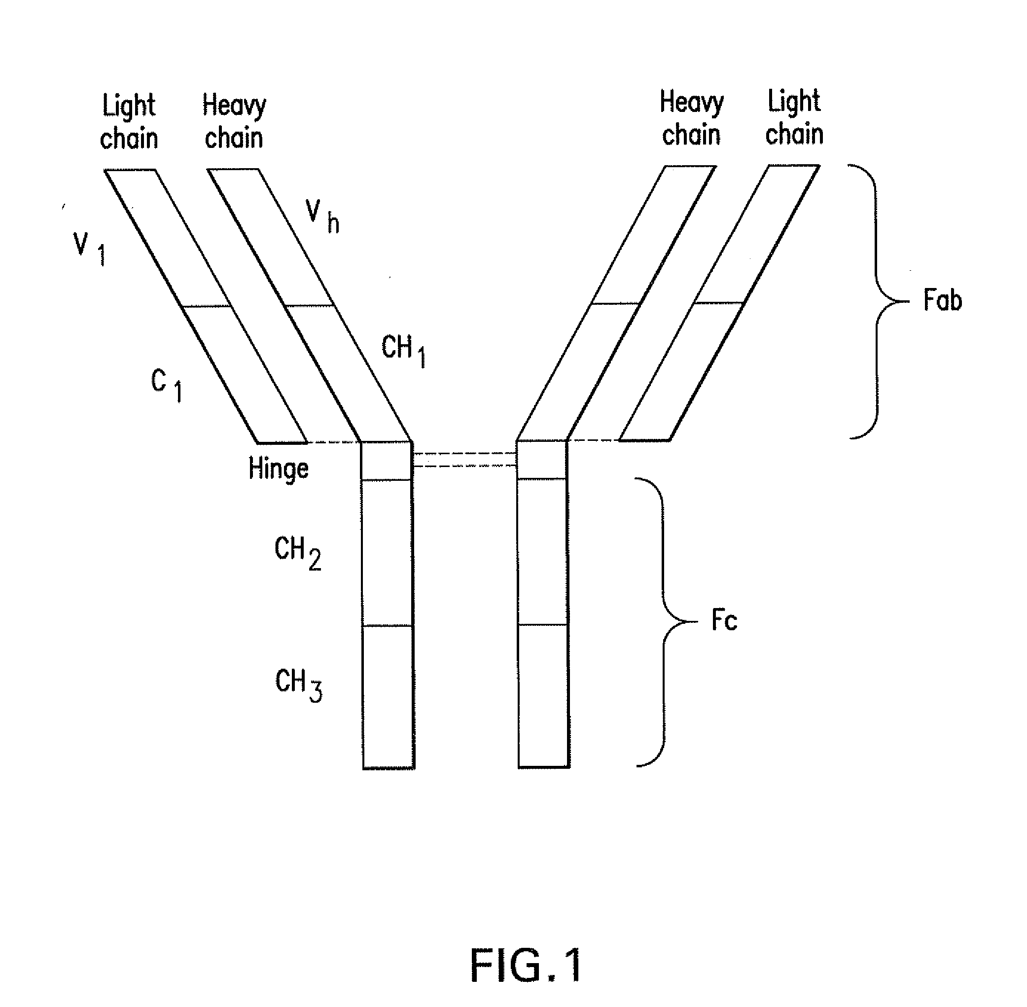
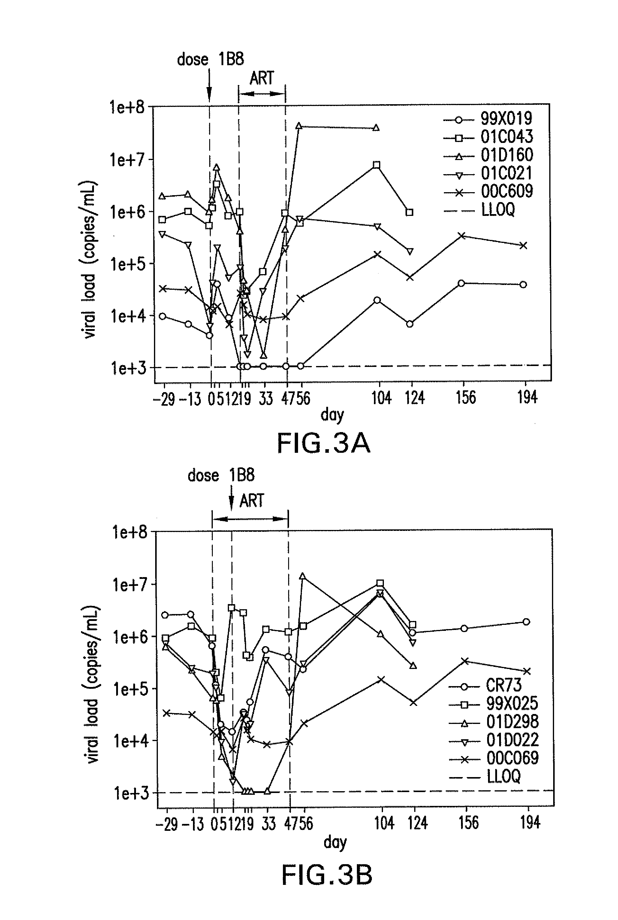
[0015]The monoclonal antibody mAb 1B8 is an immunoglobulin having two light chains comprising a variable domain (Vl) with an amino acid sequence of SEQ ID NO:5 and two heavy chains comprising a variable domain (Vh) with an amino acid sequence of SEQ ID NO:4. MAb 1B8 binds PD-1 with an especially high affinity and also blocks PD-L1 from binding to the receptor. Studies presented herein with mAb 1B8 indicate the potential of this antibody to reduce pathogen loads during persistent infection, as well as its utility to enhance T cell responsiveness to increase immunity in response to prophylactic and / or therapeutic vaccines. In particular, in a virus-infected rhesus model, administration of mAb 1B8 in combination with antiretroviral therapy resulted in an undetectable viral load in a subset of the tested animals. Vaccination of naïve rhesus macaques with an adenoviral vector vaccine containing SIV gag in conjunction with mAb 1B8 enhanced vaccine-induced T cell responses, even in the presence of host-generated antibodies against this murine antibody. As is generally appreciated by one skilled in the art, reducing the host-generated immune response to the antibody through various antibody optimization techniques described infra is expected to enhance or sustain efficacy. Antibodies in the same class as mAb 1B8 (i.e., compete for the same or proximate epitope within PD-1 as mAb 1B8, as described herein), and optimized versions thereof, will also be useful to enhance T cell responsiveness for the purposes described above.
[0026]A protein that competes with either mAb 1B8, mAb 28.11, mAb 1.8A10, mAb 1G7, mAb 20B3.1, mAb 7G3, mAb 3H4, mAb 6D10 or mAb 2.3A9 reduces binding of that monoclonal antibody to human PD-1 by at least about 20%, preferably at least about 50%, when excess and equal amounts of the competing protein and monoclonal antibody are employed.
[0037]A therapeutically effective amount is an amount sufficient to provide a useful therapeutic or prophylactic effect against a particular disease or disease condition. For example, for a patient infected with a harmful bacteria or virus, an effective amount is sufficient to achieve one or more of the following therapeutic effects: reduce the ability of the bacteria or virus to propagate in the patient or reduce the amount of bacteria or viral load in the patient. With respect to anti-infective applications for an uninfected patient, an effective amount is sufficient to achieve one or more of the following prophylactic effects: a reduced susceptibility to a bacterial or viral infection or a reduced ability of an infecting bacterium or virus to establish persistent infection. In another example, a therapeutically effective amount of a PD-1 binding protein includes an amount sufficient to generate an immune response useful to provide a therapeutic or prophylactic effect against a particular disease or disease condition. For example, for a patient with cancer, an effective amount of a PD-1 binding protein includes an amount sufficient to reduce the proliferation of cancerous cells, including the growth of tumors.

Problems solved by technology

Disruption of this inhibitory function of PD-1 can lead to autoimmunity.
The reverse scenario can also be deleterious.
Unfortunately, many viruses or microbes exploit the inhibitory functions of PD-1 to evade host T cell immunity (Greenwald et al., supra; Sharpe et al., supra).
Fifth, high degrees of PD-L1 expression in tumors (detected by immunohistochemical staining) are associated with poor prognosis for many human cancer types (Hamanishi et al., 2007, Proc. Natl. Acad. Sci.

Method used


Image

  • Pd-1 binding proteins
  • Pd-1 binding proteins
  • Pd-1 binding proteins

Examples


example 1

Generation of Monoclonal Antibodies to PD-1

[0225]Monoclonal antibodies directed to human PD-1 (SEQ ID NO:1) were generated by vaccinating mice with a DNA expression vector which encodes human PD-1.

[0226]Construction of PD-1 vector for mouse vaccination and hybridoma generation: Synthetic nucleotide gene fragments encoding amino acid sequences of human PD-1 (SEQ ID NO:1; NCBI Genbank Accession no. NP—005009) were constructed. The fragments were inserted into the V1Jns vector (SEQ ID NO:3; Shiver et al., “Immune Responses to HIV gp120 Elicited by DNA Vaccination,” in Vaccines 95, Eds. R. M. Chanock, F. Brown, H. S. Ginsberg & E. Norrby, Cold Spring Harbor, N.Y.: Cold Spring Harbor Laboratory Press, 1995, 95-98) at Bgl II site to create V1Jns-PD-1. The authenticity and orientation of the inserts were confirmed by DNA sequencing.

[0227]Production of stable cell lines: For generation of permanent cell lines, PD-1 and PD-L1 (SEQ ID NO:2; NCBI Genbank Accession no. AAP13470) fragments were ...

example 2

Biophysical Screening

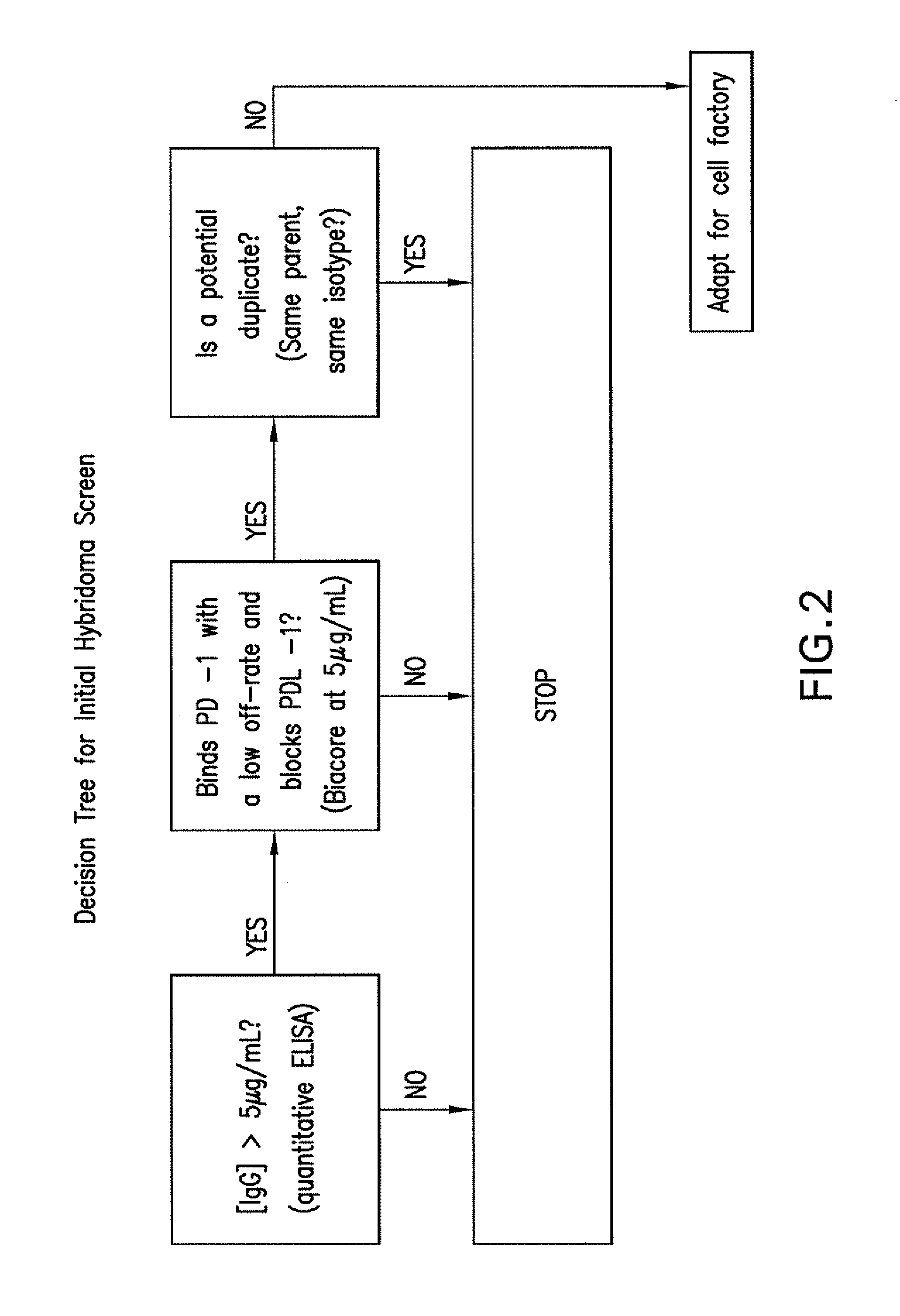
[0229]Further screening was accomplished by biophysical methods. The flowchart in FIG. 2 was followed. Concentrations of hybridoma supernatants were determined by a quantitative anti-mouse IgG ELISA, and if the concentration was greater than 5 μg / mL, supernatants were diluted to 5 μg / mL. Clones producing less than 5 μg / mL mouse IgG were not pursued.

[0230]An assay was then developed on the Biacore® platform to determine binding to PD-1, blocking of PD-L1, and to counter-screen for the human Fc portion of a recombinant PD-1 / Fc fusion protein (R&D Systems) used. Biacore® instruments incorporate microfluidics technology and surface plasmon resonance (SPR). The angle of polarized light reflecting from the side of a gold-coated sensor opposite to the solution phase is a function of the evanescent wave extending into the solution phase and sensitive to the refractive index near that interface. This refractive index is proportional to the mass density of adsorbates (e.g...

example 3

In Vitro Cell-Based Binding and Blocking Data

[0234]Several in vitro experiments were performed to determine both the binding of each identified mAb to cells expressing PD-1 and the ability to block PD-L1. 293 cells were transfected to express PD-1 on their cell membranes. Human CD4+ cells were activated by incubation with anti-CD3 / anti-CD28 beads (approximately 1 bead: 1-2 cells) for 48-72 hours at 37° C. Rhesus CD4+ cells (2×10⁶ cells / mL) were activated by incubation in plates coated with 1 μg / mL anti-CD3 / anti-CD28 for 24-72 hours at 37° C.

[0235]The results are summarized in Table 4. Criteria for further development included the following: a: binding to PD-1 on human CD4 cells (>100 relative MFI units), b: cross-reactive with PD-1 on rhesus CD4 cells (>700 relative MFI units), c: blocking of recombinant human PD-L1 / Fc binding to PD-1 expressed on transfected 293 cells (>90% inhibition), d: blocking of recombinant human PD-L1 / Fc binding to human PD-1 on activated human CD4 T cells (...


PUM

PropertyMeasurementUnit
Temperatureaaaaaaaaaa

Abstract

The present invention features PD-1 binding proteins, a subset of which inhibits binding of PD-L1 to the PD-1 receptor. These binding proteins can be employed to modulate the immune system through the manipulation of the PD-1 signaling pathway, enhancing host immunity to treat infections and cancer.

Description

CROSS-REFERENCE TO RELATED APPLICATIONS[0001]The present application claims the benefit of U.S. Provisional Application No. 61 / 069,060, filed Mar. 12, 2008, hereby incorporated by reference herein.BACKGROUND OF THE INVENTION[0002]The references cited throughout the present application are not admitted to be prior art to the claimed invention.[0003]Programmed Cell Death 1 (PD-1) is a 50-55 kDa type I transmembrane receptor originally identified by subtractive hybridization of a mouse T cell line undergoing apoptosis (Ishida et al., 1992, Embo J. 11:3887-95). A member of the CD28 gene family, PD-1 is expressed on activated T, B, and myeloid lineage cells (Greenwald et al., 2005, Annu. Rev. Immunol. 23:515-48; Sharpe et al., 2007, Nat. Immunol. 8:239-45). Human and murine PD-1 share about 60% amino acid identity with conservation of four potential N-glycosylation sites and residues that define the Ig-V domain. Two ligands for PD-1 have been identified, PD ligand 1 (PD-L1) and ligand 2 ...

Claims


Application Information

Patent Timeline
IPC IPC(8): A61K39/395C07K16/18A61P37/04
CPCC07K16/2896C07K2317/24C07K2319/30C07K2317/565C07K2317/92C07K2317/56A61P37/04
Inventor FINNEFROCK, ADAM C.FU, TONG-MINGFREED, DANIEL C.CASIMIRO, DANILO R.LI, FENGSHENGTANG, AIMIN
Owner MERCK SHARP & DOHME LLC
Learn More