
Method of treating hearing loss

Inactive Publication Date: 2010-05-27
OLESKEVICH SHARON +2
View PDF3 Cites 4 Cited by
  • Summary
  • Abstract
  • Description
  • Claims
  • Application Information

AI Technical Summary

Benefits of technology

[0044]One advantage of the present invention is that adult olfactory neuroepithelium tissue offers an abundant and easily accessible source of adult olfactory precursor cells, a significant advantage for future autotransplantation cell therapy. Olfactory receptor neurons are exposed to the external environment and are susceptible to toxic airborne chemicals, infectious pathogens and physical damage following frontal head trauma. Hence, olfactory receptor neurons are replaced periodically throughout adult life and also have the capacity to proliferate in response to acute injury. The persistence and ability of the olfactory system to regenerate its neuroepithelium by replacing damaged or dead neurons is unique in the mammalian nervous system. Replacement and proliferation is due to the presence of multipotent stem cells in the olfactory neuroepithelium (Graziadei and Graziadei, 1979; Mackay-Sim and Kittel, 1991; Roisen et al., 2001; Ortman et al., 2003; WO 03 / 064601). Importantly, the ability to obtain olfactory precursor cells from neuroepithelium in the nasal cavity without permanent damage to the donor individual eliminates the need to use highly invasive and damaging procedures. Further it provides stem cells for autologous transplantation thereby reducing both the frequency and severity of rejection.
[0045]Once removed from a subject, the olfactory neuroepithelium may be cultured to obtain adult olfactory precursor cells. For example, the minced neuroepithelium tissue can initially be placed into Dulbecco's modified eagle medium (DMEM) containing 1% (w / v) bovine serum albumin (BSA) (Sigma Chemicals, St Louis, USA), 50 μg / ml DNase (Sigma Chemicals, St. Louis, USA), 1 mg / ml hyaluronidase (Sigma Chemicals, St Louis, USA), 1 mg / ml collagenase (Roche, Australia) and 5 mg / ml dispase (Roche, Australia) for 1 hour at 37° C. The tissue suspension can subsequently be triturated, filtered through 150 μm wire mesh (Small Parts Inc., Miami Lakes, Fla., USA), centrifuged and resuspended in Neurobasal medium (Gibco BRL, MD, USA), containing 10% (w / v) dialysed fetal calf serum (FCS) (Gibco BRL, MD, USA), 10000 U / ml penicillin G (Sigma Chemicals, St. Louis, USA) and 20 mM glutamine (CSL, Melbourne, Australia). Cells can be filtered again through a 40 μm nylon mesh filter (BD Falcon, Franklin Lakes, Mass., USA) and collected on a 10 μm nylon mesh filter (Small Parts Inc, Miami Lakes, Fla., USA). Cultures may subsequently be grown at 37° C., 5% CO2 in Neurobasal medium containing B27 supplement (instead of FCS), 20 ng / ml fibroblast growth factor-2 (Promega, Madison, Wis., USA), 20 ng / ml epidermal growth factor (Promega, Madison, Wis., USA), 10000 units / ml penicillin G, and 20 mM glutamine.
[0046]Other media may be appropriate, as recognised by a person skilled in the art, as well as different animal sources of sera or the use of serum free media. Furthermore, some cultures may require additional supplements, including amino acids, growth factors etc. A variety of substrata may be used to culture the cells, for example, plastic or glass, coated or uncoated substrata may be used. For example, the culture plate may be a laminin-fibronectin coated plastic plate. Alternatively, the substrata may be coated with extracellular matrix molecules (to encourage adhesion or to control cellular differentiation), collagen or poly-L-lysine (to encourage adhesion free of biological effects). The cell culture substrata may also be treated to be charged. In the case where substratum adhesion is undesired, spinner cultures may be used, wherein cells are kept in suspension. Adult olfactory precursor cells obtained from this culturing procedure may then be used for administration to the inner ear of the subject.
[0047]The present invention also relates to the use of progeny cells which are produced from in vitro culture of adult olfactory precursor cells derived from the olfactory neuroepithelium. Adult olfactory precursor cells proliferate to form tight clusters of progeny cells referred to as neurospheres after 24 hours in culture. Generally neurospheres represent a population of neural cells in different stages of maturation formed by a single, clonally expanding precursor cell. Progeny cells that make up these neurospheres are suitable for use in the present invention.
[0048]In a preferred embodiment of the invention, human nasal mucosa is cultured to obtain a neurosphere culture (see Murrell et al., 2005). For example, human nasal mucosa biopsies may initially be placed in DMEM / HAM F12 (Invitrogen, Australia) medium supplemented with 10% FCS, penicillin and streptomycin and incubated for 45 minutes at 37° C. in a 2.4 U / ml Dispase II solution (Boehringer, Germany). Laminae propriae may be separated from the epithelium under a dissection microscope with a microspatula. Sheets of olfactory epithelium may be mechanically dissociated while lamina propriae may be cut into pieces of, for example, approximately 40 μm2 using a McElwain chopper (Brinkmann, Canada) and incubated in a 0.25 mg / ml collagenase H solution (Sigma, USA) for 10 minutes at 37° C. After mechanical trituration, the enzymatic activity may be stopped using a 0.5 mM ethylenediaminetetraacetic acid (EDTA) solution (Invitrogen, Australia). Cell pellets of both tissues may be resuspended in DMEM / HAM F12 culture medium containing 10% FCS plus penicillin / streptomycin and sequentially plated into flasks pre-treated with poly-L-lysine (1 μg / cm2; Sigma, USA). Eighteen hours after initial plating, floating cells and undigested pieces of epithelium and lamina propria may be transferred to other coated wells. This operation may be repeated 24 hours later.
[0049]According to the invention, the adult olfactory precursor cells or progeny thereof may be collected from culture and resuspended in a buffer before being administered to the inner ear of the subject. In one embodiment of the invention, the adult olfactory cells or progeny thereof are harvested and frozen in, for example, serum / 10% DMSO prior to resuspension in the buffer. A variety of methods may be used to collect adult olfactory precursor cells or progeny thereof, including enzymatic removal (such as by trypsination), chemical methods, (e.g. cation metal chelation using EDTA or ethylene glycol-bis(β3-aminoethyl ether) NNN′N′-tetraacetic acid (EGTA), and mechanically, such as by cell scraping or in the case of cell suspension, by simple centrifugation. The “buffer” may be any suitable buffer that is generally regarded as safe and generally confers a pH from or about 4.8 to 8, preferably from or about 5 to 7. Examples include acetic acid salt buffer, which is any salt of acetic acid, including sodium acetate and potassium acetate, succinate buffer, phosphate buffer, citrate buffer, histidine buffer, or any others known in the art to have the desired effect.Implantation of Adult Olfactory Precursor Cells into the Inner Ear

Problems solved by technology

There is evidence of regeneration of sensory cells in non-mammalian species (Stone and Rubel, 2000; Morest and Cotanche, 2004), however, studies in mammals have failed to find evidence of cochlear regeneration (Forge and Nevill, 1998).
The potential for stem cell therapy for treatment of sensorineural hearing loss in humans, in particular the use of stem cells originating from human embryos or aborted foetal tissues is complicated by moral and ethical considerations.
While the possibility of deriving stem cells from adult tissue sources holds real promise, there are also some significant limitations.
First, adult stem cells are often present in minute quantities, are difficult to isolate and purify, and their number may decrease with age.
Further, although stem cells have been isolated from the adult central nervous system, it is not currently possible to remove them without serious consequences to the donor.

Method used


Image

  • Method of treating hearing loss
  • Method of treating hearing loss
  • Method of treating hearing loss

Examples


example 1

Materials and Methods

Primary Adult Olfactory Precursor Cells

[0064]To isolate olfactory precursor cells, olfactory turbinates were dissected from 10-15 adult CBA / CaH mice aged 6 weeks. Mice were anaesthetised with CO2, decapitated and olfactory turbinates were removed and placed in DMEM containing 9.6 mg / ml HEPES buffer. Tissue was centrifuged and supernatant was removed before placing minced tissue into DMEM containing 1% (w / v) BSA (Sigma Chemicals, St Louis, USA), 50 μg / ml DNase (Sigma Chemicals, St. Louis, USA), 1 mg / ml hyaluronidase (Sigma Chemicals, St. Louis, USA), 1 mg / ml collagenase (Roche, Australia) and 5 mg / ml dispase (Roche, Australia) for 1 hour at 37° C. Tissue suspension was triturated, filtered through a 150 μm wire mesh (Small Parts Inc., Miami, Fla., USA), centrifuged and resuspended in Neurobasal medium (Gibco BRL, MD, USA), containing 10% (w / v) dialysed FCS (Gibco BRL, MD, USA), 10000 U / ml Penicillin G (Sigma Chemicals, St. Louis, USA) and 20 mM glutamine (CSL, Me...

example 2

Characterisation of Primary Olfactory Precursor Cells

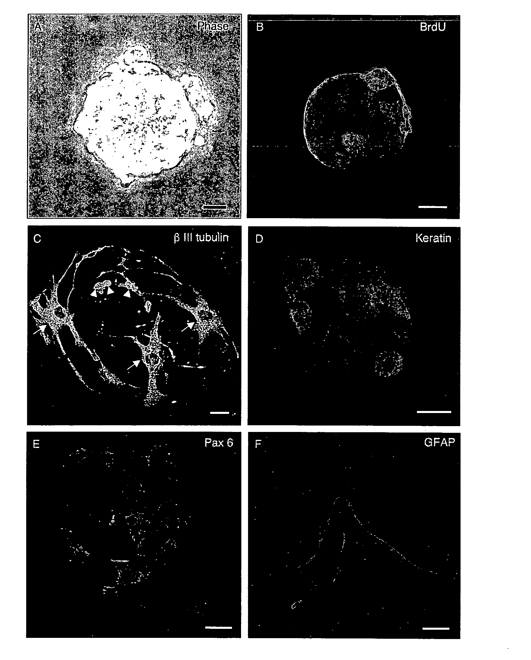
[0082]Olfactory precursor cells were prepared from dissociated primary cell cultures (n=16) of adult mouse olfactory turbinates. Precursor cells proliferated to form tight clusters of cells (neurospheres) after 24 hours in culture. Neurospheres were typically 100-200 μm in diameter, but could reach 500 μm in diameter (see FIG. 1A). Single round cells radiating from the neurospheres were commonly seen (see FIG. 1A). Some cells within the neurospheres were immunopositive for the BrdU antibody, a marker of the S-phase of the cell-cycle (see FIG. 1B). Trituration of neurospheres and single cell disposition experiments using fluorescence-activated cell sorting (FACS Calibur, BD Falcon, Franklin Lakes, Mass., USA) gave rise to secondary neurospheres, which provides additional evidence that neurospheres were self-replicating (data not shown). Upon removal of growth factors, olfactory neurospheres spontaneously differentiated into cells s...

example 3

Characterisation of Hair Cells in the Organ of Corti

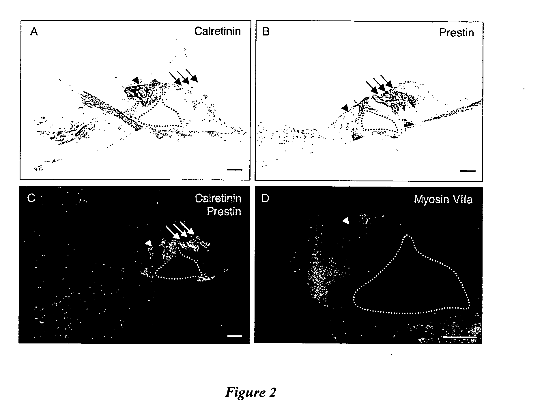
[0083]Hair cells in the adult organ of Corti (cochlea, n=9) were analysed in thin sections of cochlea using markers of inner and outer hair cells. Inner hair cells and spiral ganglion nerve fibres showed positive immunoreactivity to the calcium binding protein, calretinin (see FIG. 2A). No staining was observed in outer hair cells with the calretinin antibody. Outer hair cells were positively labelled with prestin, a transmembrane motor protein present in these cells (see FIG. 2B). Double labelling immunofluorescence shows there was no co-localisation of calretinin and prestin in the adult mouse organ of Corti, confirming the specificity of these antibody markers (see FIG. 2C). Myosin VIIa, a motor protein essential to sensory epithelia, labelled the cytoplasm of the inner hair cell shown in FIG. 2D.


PUM


Abstract

The present invention relates to a method of generating cochlear hair cell-like cells in the inner ear of a subject. Preferably, the subject is human and is suffering from sensorineural hearing loss. The invention also relates to a method of generating cochlear hair cell-like cells in vitro and use of the cochlear hair cell-like cells for treating or preventing sensorineural hair loss in a subject.

Description

FIELD OF THE INVENTION[0001]The present invention relates to methods of generating cochlear hair cell-like cells in vivo and in vitro. The methods of the invention are useful in the treatment of inner hair cell related conditions, for example, sensorineural hearing loss.BACKGROUND OF THE INVENTION[0002]Sensorineural deafness induced by ageing, excessive noise and certain antibiotics accounts for the majority of permanent hearing loss in humans. Hearing loss is caused by the dysfunction of the sensory epithelium (the organ of Corti) within the inner ear (cochlea). It is associated with the irreversible loss of sensory hair cells (Raphael, 2002) and spiral ganglion neurons (Raphael, 2002). There is evidence of regeneration of sensory cells in non-mammalian species (Stone and Rubel, 2000; Morest and Cotanche, 2004), however, studies in mammals have failed to find evidence of cochlear regeneration (Forge and Nevill, 1998).[0003]Several areas of research have addressed the treatment of s...

Claims


Application Information

Patent Timeline
IPC IPC(8): A61K35/12C12N5/00A61P27/16A61K35/30
CPCA61K35/30A61P27/16
Inventor OLESKEVICH, SHARONDOYLE, KHARENMCKAY, SARAH
Owner OLESKEVICH SHARON
Learn More