
Biocompatible Coated Nanostructured Titanium Surfaces

a nano-structured titanium and biocompatible technology, applied in the field of biomaterials, can solve the problems of insufficient practical use, unpredictable cell/tissue adhesion, and long-term failure of titanium implants, and achieve the effect of strengthening bone growth and strong cell adhesion

Inactive Publication Date: 2010-02-04
METASCAPE
View PDF9 Cites 80 Cited by
  • Summary
  • Abstract
  • Description
  • Claims
  • Application Information

AI Technical Summary

Benefits of technology

[0021]Accordingly, whether immobilized inside or outside the nanotubes, biomaterials can be released in a time-dependent fashion in vivo. This is particularly attractive for use in various implants and becomes possible because the biomaterials are not covalently attached to the substrate surface. Nanotube features can be designed so that adherence of the coating can be modified to be relatively weak or, by adjusting the size of the tubes, change the ratio of bioactive material inside and outside the tubes.
[0023]When used as coatings on implants, the biomolecules act not only as anchors for osteoblasts but also enhance bone growth in vivo. As demonstrated herein, the RGD tripeptide sequence was deposited on a nanostructured Ti surface and showed strong cell adhesion for osteoblasts and fibroblasts.

Problems solved by technology

However, the normal oxide layer of titanium is not sufficiently bioactive to form a direct bond with juxtaposed bone, which may translate into a lack of osseointegration, leading to long-term failure of titanium implants.
A disadvantage of these approaches is that neither the mechanical nor the chemical methods produce highly controllable topological properties, and cell / tissue adherence may be unpredictable or insufficient for practical use.
In some cases, the methods may cause formation of surface residuals, which can be interfere with osteoblast (bone forming cell) adherence and function.
The peptide compositions were administered to the site where mineralization was desired; however, there was no evidence that these or other polypeptides immobilized on appropriate substrates could be used as effective scaffolds to promote bone mineralization.
Despite studies demonstrating that peptide-modified surfaces influence in vitro cellular behavior, it is recognized that in vivo use may raise issues of undesirable proteolysis and that manufacturing costs for the bound peptides may be high.
Despite progress in modifying metal surfaces to improve tissue and cell adhesion properties, adequate in vivo osseointegration on implant prostheses remains a challenge.

Method used


Image

  • Biocompatible Coated Nanostructured Titanium Surfaces
  • Biocompatible Coated Nanostructured Titanium Surfaces
  • Biocompatible Coated Nanostructured Titanium Surfaces

Examples


example 1

Anodized Titanium

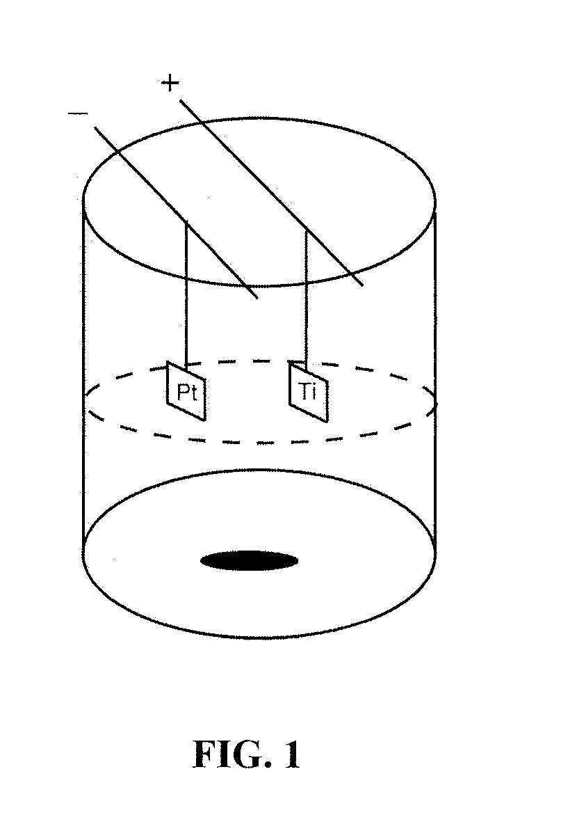
[0089]A standard anodization apparatus utilizing a platinum cathode and titanium anode connected by copper rods to a power supply was employed (FIG. 1). The beaker is Teflon® or other material impervious to the acid.

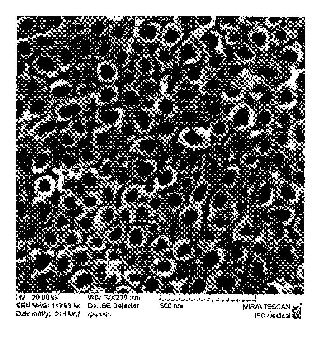
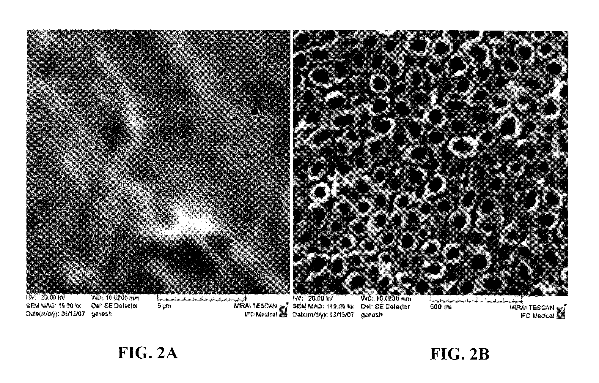
[0090]Rectangular shaped titanium foil with a thickness of 250 μm (99.7%; Alfa Aesar) was cleaned ultrasonically with ethanol and water prior to anodization. Cleaned substrates were etched with a mixture of 1M HNO3 (Aldrich) (with few drops of HF solution) and further cleaned with deionized water. Afterwards, pretreated specimens were anodized in a 1.5% hydrofluoric acid (HF) solution. Using a DC power supply, a 20 V anodizing voltage was applied for 10 minutes. During processing, the anode and cathode were kept parallel with a separation distance of about 1 cm. Specimens were rinsed with deionized water and dried with nitrogen gas immediately after being anodized. Before osteoblast adhesion was performed, specimens were sterilized under UV for 1 hr in a...

example 2

Molecular Plasma Deposition Apparatus

[0102]The deposition apparatus includes a vacuum chamber with a small aperture, and a small bore, metallic needle connected to a tube connected to a reservoir holding a liquid suspension or solution of the material desired to be deposited. The reservoir is at atmospheric pressure. A power supply with the ability to supply up to 60 kV can be employed; however, the voltage attached to the needle is typically −5000 volts to +5000 volts. A substrate inside the vacuum chamber, is centered on the aperture with a bias from −60 kV through −60 kV, including ground. The apparatus is illustrated in FIG. 3A.

[0103]Another molecular deposition apparatus is illustrated in FIG. 3B. This is a modification of the apparatus in FIG. 3A such that the needle, tube, and reservoir are disposed in an enclosure that excludes air, but allows for the controlled introduction of other gases. Optionally selected gases include argon, oxygen, nitrogen, xenon, hydrogen, krypton, ...

example 3

Osteoblast Cell Adhesion to Anodized Titanium

[0105]Human osteoblasts (CRL-11372 American Type Culture Collection, population numbers 7-8) in Dulbecco Modified Eagle Medium (Gibco) supplemented with 10% fetal bovine serum (Hyclone) and 1% Penicillin / Streptomycin (Hyclone) were seeded at a density of 3500 cells / cm2 onto an anodized titanium substrate and placed in standard cell culture conditions (humidified, 5% CO2 / 95% air environment) for 4 hr. The substrate was rinsed in phosphate buffered saline to remove any nonadherent cells. The remaining cells were then fixed with formaldehyde (Aldrich Chemical Company, USA), stained with Hoescht 33258 dye (Sigma), and counted under a fluorescence microscope (Leica, DM IRB). Five random fields were counted per sample substrate. All tests were run in triplicate and repeated at least three separate times. Standard t-tests were used to check statistical significance between means.

[0106]Results showed significantly increased (p<0.01) osteoblast ad...


PUM

PropertyMeasurementUnit
Diameteraaaaaaaaaa
Diameteraaaaaaaaaa
Diameteraaaaaaaaaa

Abstract

Bioactive molecules have been coated on nanotubular structured titanium substrates by molecular plasma deposition. The coatings promote cell adhesion and are particularly suited for orthopedic implants that provide improved bone cell adhesion and new tissue growth. Nanodimensional features on titanium substrates are engineered using electrochemical anodization techniques. The nanostructured surfaces provide superior support for a wide selection of polypeptide coatings.

Description

[0001]This application claims benefit of U.S. Provisional Application Ser. No. 60 / 934,279 filed Jun. 12, 2007, which is hereby incorporated by reference herein in its entirety, including any figures, tables, nucleic acid sequences, amino acid sequences, or drawings.BACKGROUND OF THE INVENTION[0002]1. Field of the Invention[0003]The invention relates generally to biomaterials and particularly to bioactive protein-coated nanostructured titanium substrates.[0004]2. Description of Background Art[0005]Titanium and its alloys have been widely used to create dental and orthopedic implants because of their excellent biocompatibility and mechanical properties. Titanium (Ti) spontaneously forms an oxide layer up to a thickness of about 2 to 5 nm both in air and in the body, providing corrosion resistance. However, the normal oxide layer of titanium is not sufficiently bioactive to form a direct bond with juxtaposed bone, which may translate into a lack of osseointegration, leading to long-ter...

Claims


Application Information

Patent Timeline
IPC IPC(8): A61K9/00A61K38/02A61K38/07A61K38/08A61K38/16A61K38/14C12N5/00A61K35/12A61P43/00
CPCA61L27/06A61L27/34A61L27/54A61L2300/25A61L2300/252A61L2300/412C07K17/14A61L2300/606A61L2400/12C08L89/00A61P43/00
Inventor BALASUNDARAM, GANESANSHIMPI, TUSHAR M.STOREY, DANIEL M.
Owner METASCAPE
Learn More