
Purification and protective efficacy of monodisperse and modified yersinia pestis capsular f1-v antigen fusion proteins for vaccination against plague

a technology of capsular f1-v and yersinia pestis, which is applied in the field of compounding and methods, can solve the problems of low process yield, common side effects, and failure to fully realize the development of vaccines using only fl antigens, and achieve the effect of inhibiting yersinia pestis infection

Inactive Publication Date: 2009-05-21
THE UNITED STATES OF AMERICA AS REPRESENTED BY THE SECRETARY DEPARTMENT OF HEALTH AND HUMAN SERVICES NATIONAL INSTITUTES OF HEALTH
View PDF0 Cites 7 Cited by
  • Summary
  • Abstract
  • Description
  • Claims
  • Application Information

AI Technical Summary

Benefits of technology

[0008]Disclosed herein is an improved F1-V vaccine that includes a substantially monodisperse immunogenic F1-V fusion protein. Unlike the previous F1-V fusion protein vaccine, the fusion protein described herein is substantially monomeric and does not tend to self-associate and form aggregates, yet it retains its immunogenicity.
[0009]Also disclosed are pharmaceutical compositions that include the substantially monodisperse immunogenic proteins as well as methods for eliciting an immune response in a subject, which methods include (a) selecting a subject in which an immune response to the substantially monodisperse immunogenic F1-V protein is desirable; and (b) administering to the subject...

Problems solved by technology

The whole-cell killed vaccine previously was available for people at possible high risk of exposure, such as military or laboratory personnel, but side effects were common, and multiple boosters were necessary.
However, attempts to develop a vaccine using only the Fl antigen were less than fully successful (Friedlander et al.
Thus, the F1-V vaccine presented risks for large-scale manufacture including: 1) possible entrapment of contaminants within multimeric forms, which can lower process yields and increase process costs to achieve purity; and 2) uncontrolled or premature re-folding that can affect fusion-protein structure and thereby impact product consistency and long-term stability (Chi et al., (2003) Pharm. Res. 20, 1325-1336).

Method used


Image

  • Purification and protective efficacy of monodisperse and modified yersinia pestis capsular f1-v antigen fusion proteins for vaccination against plague
  • Purification and protective efficacy of monodisperse and modified yersinia pestis capsular f1-v antigen fusion proteins for vaccination against plague
  • Purification and protective efficacy of monodisperse and modified yersinia pestis capsular f1-v antigen fusion proteins for vaccination against plague

Examples


example 1

Materials and Methods

[0162]This Example describes materials and methods that were used in performing Examples 2-16, below. Although particular methods are described, one of skill in the art will understand that other, similar methods also can be used.

Bacterial Strain, Plasmid Construction, Cultivation, and Induction

[0163]For F1-VMN the E. coli strain BLR130 and the F1-V expression vector, pPW731 (USAMRIID), controlled under a T7 promoter, were used for F1-V expression (Powell et al., (2005) Biotechnol. Prog. 21 (2005), pp. 1490-1510). A growth medium of soytone, yeast extract, and glucose (J. T. Baker, Phillipsburg, N.J.) and the antibiotics kanamycin (30 mg / L) and tetracycline (15 mg / L; Sigma, St. Louis, Mo.) were used in phosphate buffer, pH ˜7.3. Sterile medium in shaker flasks (300 ml) was inoculated with 1 ml of the strain from a previously made glycerol stock and incubated for ˜13 hours at 37° C. with shaking at 220 rpm. Batch cultivations were carried out in a Bioflo 4500 (Ne...

example 2

F1-VMN and F1-VC424S-MN Expression

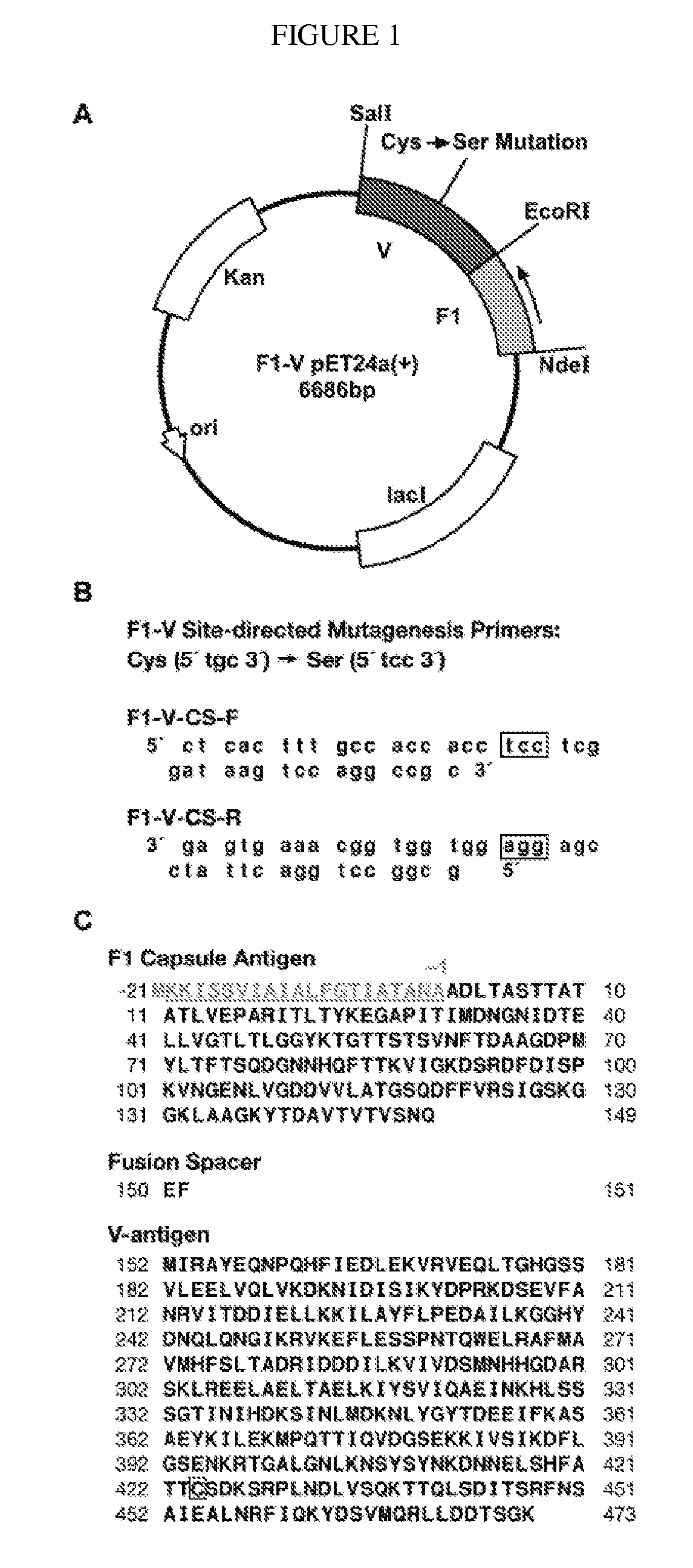
[0184]This Example demonstrates the expression of two F1-V fusion proteins, F1-VMN and F1-VC424S-MN. In order to evaluate the effect of super molecular structure (for instance, its state of aggregation) of the F1-V-based plague vaccine antigen on protective efficacy and to facilitate vaccine production, the sole cysteine (C424) in F1-V was replaced with a serine residue by site-directed mutagenesis. Standard F1-VMN and the modified F1-VC424S-MN proteins were independently over-expressed in E. coli, recovered by mechanical lysis / pH-modulation, and purified from urea-solubilized, soft inclusion bodies with successive ion-exchange, ceramic hydroxyapatite, and size-exclusion chromatography stages as described in Example 1. Aggregation characteristics for the purified proteins were characterized and compared under variable pH and buffer solution-additive conditions. The biological activities of the two purified proteins in various super molecular states ...

example 3

Ion-Exchange Chromatography

[0187]This Example demonstrates the purification of both F1-V and the variant F1-VC424S. Standard F1-V and the F1-VC424S variant purified similarly through the ion-exchange stages of the improved process. Profiles from purification of the standard (cysteine-containing) F1-V performed at the larger scale are shown (FIG. 4). The Q-Sepharose FF chromatography stage, performed under partially dissociating conditions and eluted with the denaturing Gdn cation, was effective as a charge-based step for isolating F1-V (FIG. 4A). F1-VC424S monomer eluted at Gdn HCl concentrations below 100 mM, similar to what was observed with F1-VSTD-MN. Dimer and trimer forms of F1-V remained intact during reducing SDS-PAGE analysis when samples were prepared by heating to less than 70° C. for 10 minutes. These same forms were dispersed and ran as apparent F1-V monomers when heated to 100° C. for 10 minutes, corroborating prior observations of strong self association F1-V by gel e...


PUM

PropertyMeasurementUnit
Fractionaaaaaaaaaa
Fractionaaaaaaaaaa
Molar densityaaaaaaaaaa

Abstract

This disclosure concerns compositions and methods for the treatment and inhibition of infectious disease, particularly bubonic and pneumonic plague. In certain embodiments, the disclosure concerns immunogenic proteins, for instance substantially monodisperse F1-V fusion proteins, that are useful for inducing protective immunity against Y. pestis.

Description

FIELD OF THE DISCLOSURE[0001]This disclosure concerns compositions and methods for the treatment and inhibition of infectious disease, particularly bubonic and pneumonic plague. In certain embodiments, the disclosure concerns immunogenic proteins, for instance monodisperse F1-V fusion proteins, that can be used to induce protective immunity against Y. pestis infection.BACKGROUND[0002]Plague is an infectious disease caused by the bacteria Yersinia pestis, which is a non-motile, slow-growing facultative organism in the family Enterobacteriacea. Y. pestis is carried by rodents, particularly rats, and in the fleas that feed on them. Other animals and humans usually contract the bacteria directly from rodent or flea bites.[0003]Yersinia pestis is found in animals throughout the world, most commonly in rats but occasionally in other wild animals, such as prairie dogs. Most cases of human plague are caused by bites of infected animals or the infected fleas that feed on them. Y. pestis can ...

Claims


Application Information

Patent Timeline
IPC IPC(8): A61K39/00C07K16/00C07H21/04C07K1/14C12N15/00
CPCA61K39/025A61K2039/55505C07K2319/40C07K14/24C07H21/04Y02A50/30
Inventor NELLIS, DAVID F.GIARDINA, STEVEN L.GOODIN, JEREMY
Owner THE UNITED STATES OF AMERICA AS REPRESENTED BY THE SECRETARY DEPARTMENT OF HEALTH AND HUMAN SERVICES NATIONAL INSTITUTES OF HEALTH
Learn More