
Method for modifying a medical implant surface for promoting tissue growth

a technology of medical implants and tissue growth, applied in the field of modifying the surface of medical implants to promote tissue growth, can solve the problems of recurrent cerebrovascular events, increased risk of future pfo and paradoxical embolism patients, and potential adverse side effects, and achieves the effect of reducing the risk of thrombosis and avoiding excessive fibrosis

Inactive Publication Date: 2008-04-17
WL GORE & ASSOC INC
View PDF16 Cites 92 Cited by
  • Summary
  • Abstract
  • Description
  • Claims
  • Application Information

AI Technical Summary

Benefits of technology

[0008] The invention describes configuring the surface of the septal occluder so that host tissue grows over the device, healing the patient's defect without excessive fibrosis or elevated risk of thrombosis.

Problems solved by technology

Under certain conditions, however, RA pressure can exceed LA pressure, creating the possibility for right to left shunting of blood, permitting blood clots to enter the systemic circulation.
In addition, there is evidence that patients with PFO and paradoxical embolism are at increased risk for future, recurrent cerebrovascular events.
These patients are commonly treated with oral anticoagulants, which have potential adverse side effects, such as hemorrhaging, hematoma, and interactions with a variety of other drugs.
However, currently available designs of septal closure devices present drawbacks, such as technically complex implantation procedures, high complication rates (for example, thrombi, device fractures, conduction system disturbances, perforations, and residual leaks), a high septal profile, and presentation of large masses of foreign material.
In addition, since many septal closure devices were originally designed to close ASDs, which are true holes, rather than the flap-like anatomy of most PFOs, many closure devices lack the anatomic conformability to effectively close a PFO.
In addition, some septal closure devices are complex to manufacture, which can result in lack of consistency in product performance.

Method used


Image

  • Method for modifying a medical implant surface for promoting tissue growth
  • Method for modifying a medical implant surface for promoting tissue growth
  • Method for modifying a medical implant surface for promoting tissue growth

Examples


example 1

Surface Modification Through Plasma Treatment

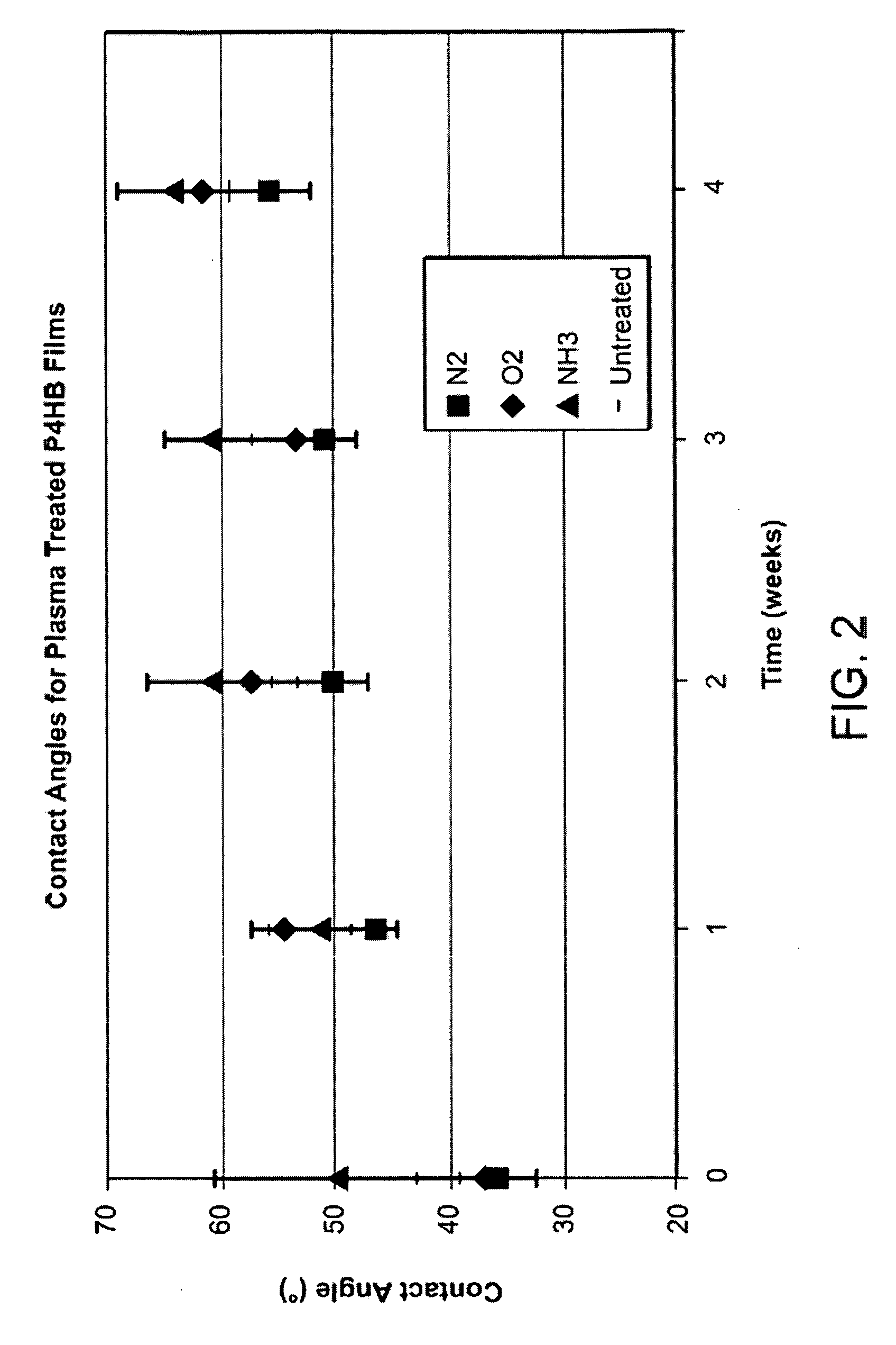
[0049] Plasma treating the surface of a septal occluder is one way to alter the surface characteristics of the material to promote protein deposition and cell attachment. Plasma treating the septal occluder increases the wetability of the implant surface, thereby improving endothelial cell attachment to the implant.

[0050] Plasma is partially ionized gas generated by applying an electrical field to a gas under at least partial vacuum. Plasma reacts and combines with first few atomic layers of the surface while the visual and bulk properties of the material remain unchanged. Gases such as oxygen and nitrogen have been used during plasma treatment, as well as gases containing amine groups.

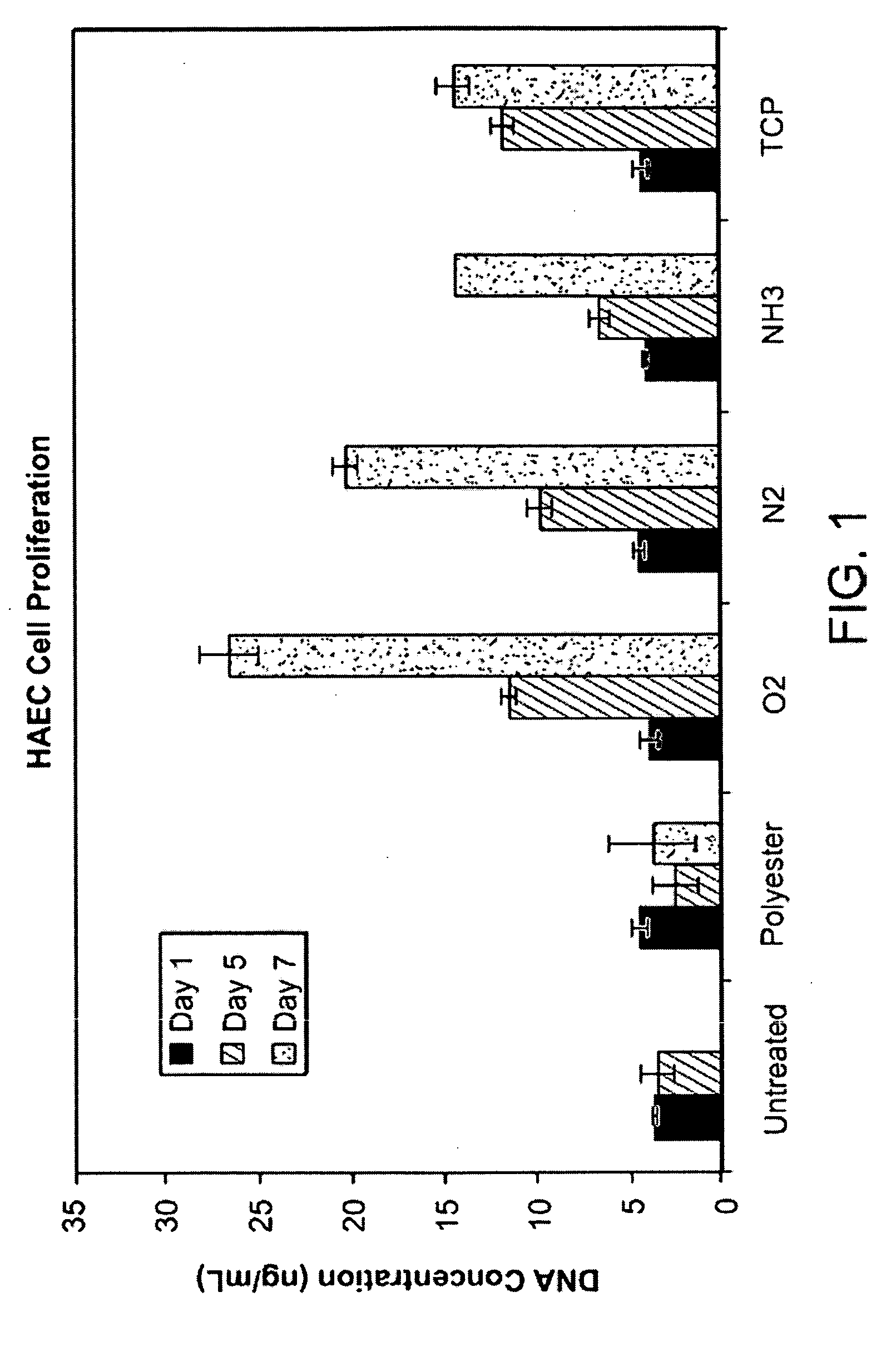
[0051]FIG. 1 is a bar graph showing the effects in vitro of plasma treatment on proliferation of HAEC cells (human aortic endothelial cells) on an untreated polyester scaffold typically used in a septal occluder (“Polyester”), an untreated bioabsorbable ...

example 2

Stability of Plasma Treated Bioabsorbable Polymers

[0054] In order to determine the stability of plasma treated P4HB, molecular weight data of plasma treated solvent cast (porous cast) P4HB samples was taken at 4 days after plasma treatment and 5 weeks after plasma treatment.

[0055] Plasma treated samples were processed at PLASMAtech (Erlanger, Ky.). Samples of P4HB were plasma treated with oxygen gas (O2), nitrogen gas (N2), nitrous oxide (N2O), and a combination of ammonia gas (NH3) and oxygen gas (O2). The molecular weight data, shown in FIGS. 3A-B, indicates that the decrease in molecular weight on a percentage basis was least for the combination NH3 / O2 treated P4HB. Accordingly, the NH3 / O2 plasma treated P4HB has the greatest stability of the plasma treatments tested. According to the invention, the ratio of NH3 to O2 used to treat the P4HB is 2:3 in one embodiment, 1:1 in another embodiment, and 1:2 in yet another embodiment.

example 3

Surface Modification by Collagen Coating

[0056] Collagen can be made recombinantly in highly purified form, free of contamination from disease-causing pathogens such as viruses and prions. It is also available commercially (e.g., from FibroGen, Inc., South San Francisco, Calif., USA). Collagen type I and type III promote tissue growth, and can be applied to the surface of a medical implant through a simple dip coating process. For example, to coat a P4HB scaffold or frame with collagen according to the invention a 40 microgram / mL solution of collagen is made by combining 0.5 mL liquid collagen with 37.5 mL PBS. The scaffold or frame is then cleaned with ethyl alcohol and deionized water prior to being soaked in the collagen solution for 15 minutes. The scaffold or frame is dried for one hour between coats. Any number of coats of collagen may be applied. Four (4) coats of collagen are optimal according to one embodiment.

[0057] Attachment of HAEC cells to variously-treated scaffolds ...


PUM

PropertyMeasurementUnit
Bioabsorbableaaaaaaaaaa

Abstract

Disclosed is an occluder for closing an intracardiac defect, such as a patent foramen ovale (PFO), and a method for making the same. The occluder includes a frame and at least one scaffold which are formed from a bioabsorbable polymer, such as poly-4-hydroxybutyrate. The surface of the frame and scaffold are textured to promote cell attachment. Texturing of the surface can be achieved by any number of mechanical or chemical procedures. The device is coated with collagen and heparin which are covalently bound to the surface of the device. The occluder provides improved defect closure compared to other septal occluders known in the art. In particular, the occluder described is specifically designed to improve host cell attachment to and tissue ingrowth over the device when implanted in a patient as compared to the level of host cell attachment and tissue ingrowth achieved with other implantable devices made of bioabsorbable polymers.

Description

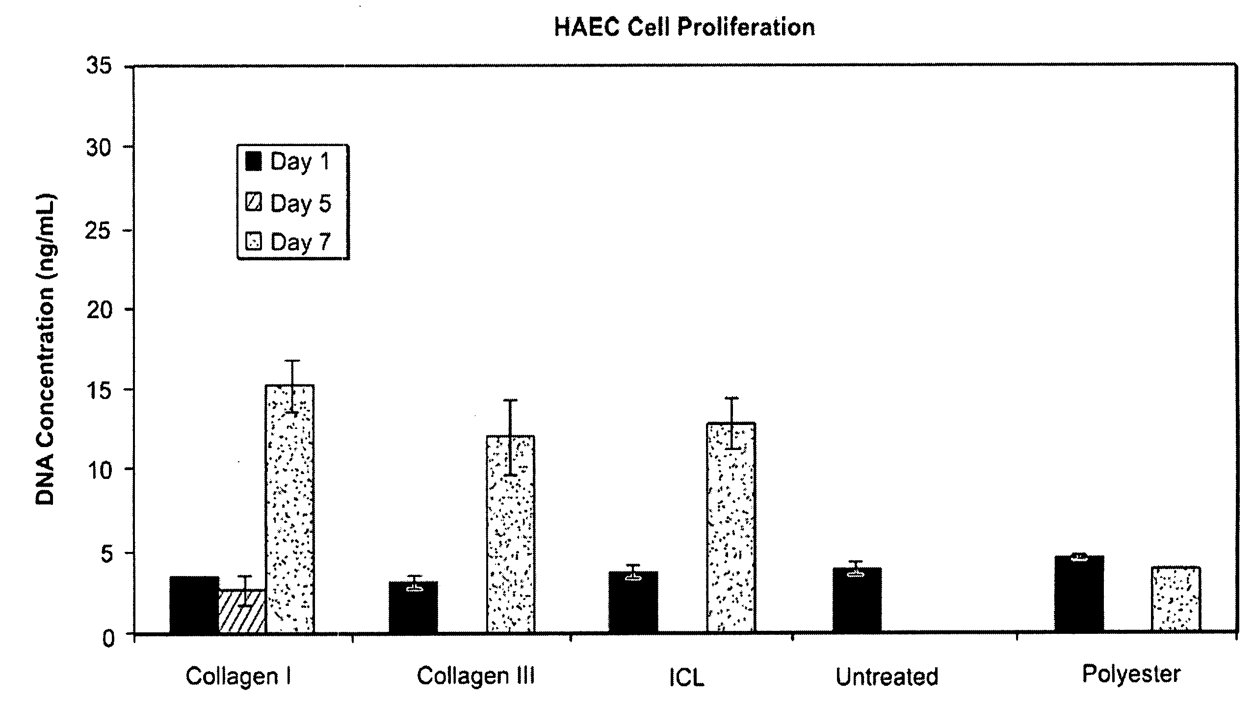
CROSS-REFERENCE TO RELATED APPLICATIONS [0001] This application claims priority to and the benefit of U.S. Provisional Patent Application No. 60 / 847,310, filed Sep. 26, 2006, the contents of which are incorporated by reference herein.BACKGROUND OF THE INVENTION [0002] A patent foramen ovale (PFO) is a persistent, one-way, usually flap-like opening in the wall between the right atrium and left atrium of the heart. Since left atrial (LA) pressure is normally higher than right atrial (RA) pressure, the flap typically stays closed. Under certain conditions, however, RA pressure can exceed LA pressure, creating the possibility for right to left shunting of blood, permitting blood clots to enter the systemic circulation. In utero, the foramen ovale serves as a physiologic conduit for right-to-left shunting. After birth, with the establishment of pulmonary circulation, the increased left atrial blood flow and pressure results in functional closure of the foramen ovale. This functional clos...

Claims


Application Information

Patent Timeline
IPC IPC(8): A61B17/03B29C59/00
CPCA61B17/0057A61B2017/00004A61B2017/00243A61L27/18A61L27/34A61L27/50B29C59/14C08L67/04C08L89/06C08L5/10
Inventor KLADAKIS, STEPHANIE M.
Owner WL GORE & ASSOC INC
Learn More