
Mutants of green fluorescent protein

a technology of green fluorescent protein and mutants, which is applied in the field of molecular and cellular biology, can solve the problems of only useful type of technique, inability to guarantee that a majority (or even any) of the target cells will take up and/or express exogenous dna, and reporter genes typically do not confer any particular advantage to the recipient, etc., and achieves the effect of being convenient to us

Inactive Publication Date: 2007-01-25
INVITROGEN
View PDF2 Cites 4 Cited by
  • Summary
  • Abstract
  • Description
  • Claims
  • Application Information

AI Technical Summary

Benefits of technology

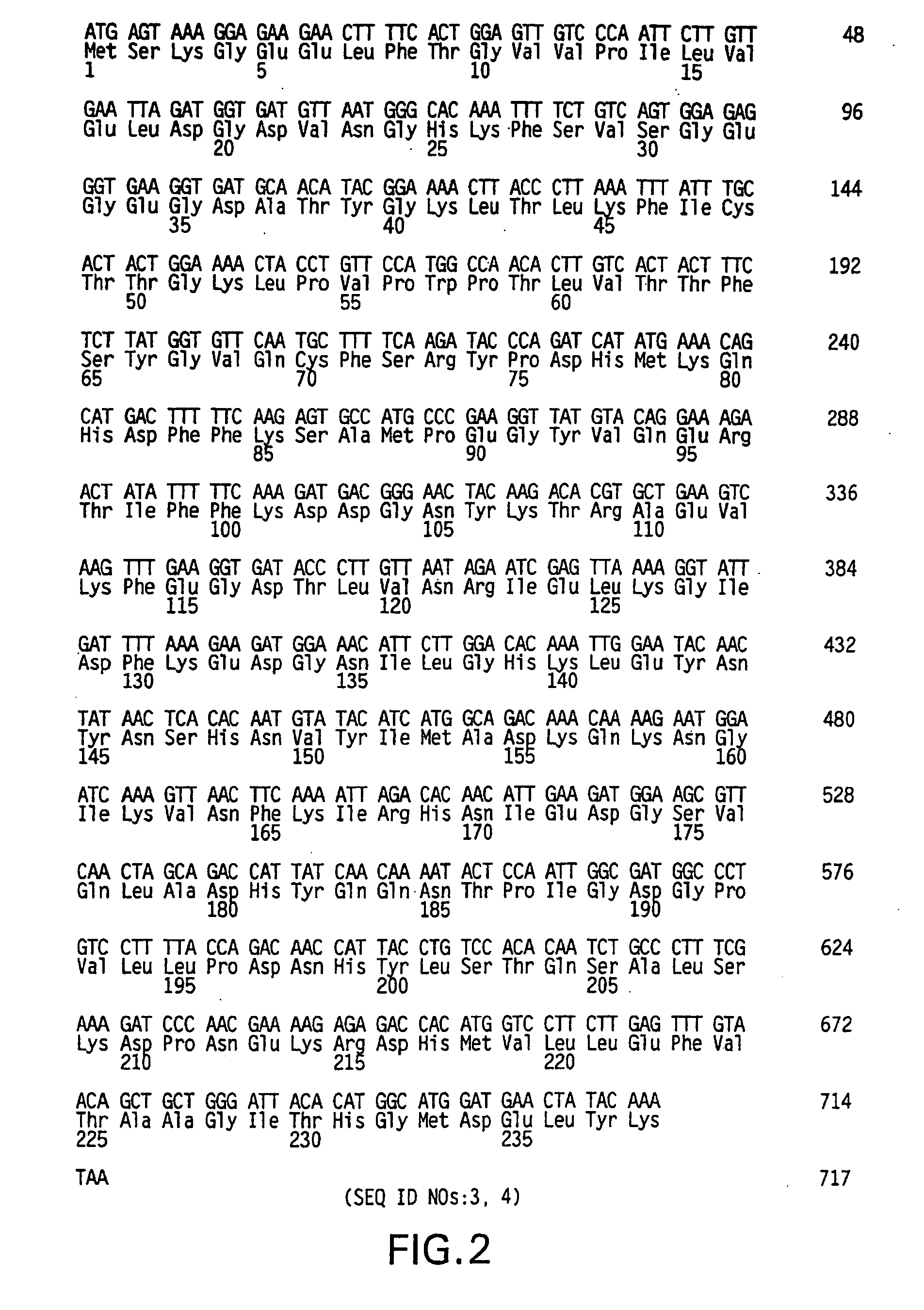
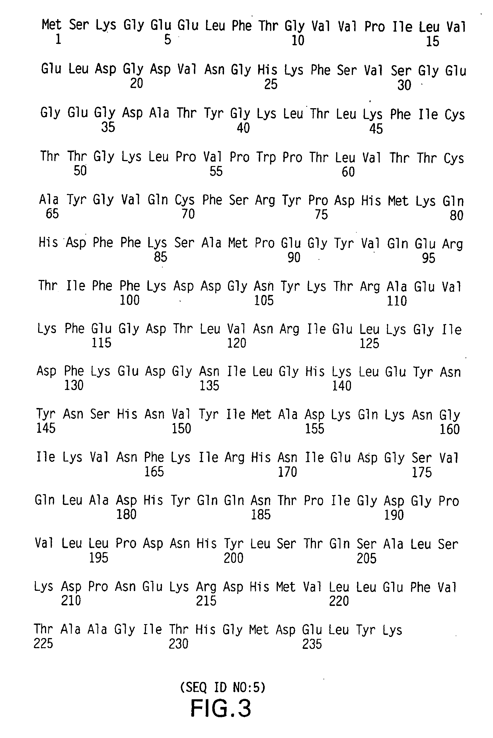
[0028] It is thus an object of the present invention to provide mutant GFP cDNAs and proteins. In one aspect, the invention relates to such mutant GFP cDNAs which, when transfected into prokaryotic (e.g., bacterial) or eukaryotic (e.g., mammalian) cells, increase the sensitivity of detection (measured as percentage or number of positive cells). The present invention thus provides nucleic acid molecules encoding mutant GFPs, wherein the mutant GFPs have an amino acid sequence comprising an amino acid residue lacking an aromatic ring structure at position 64 and an amino acid residue having a side chain no longer than two carbon atoms in length at position 65. Preferably, (a) if the residue at position 64 is leucine then the residue at position 65 is not cysteine or threonine; (b) if the residue at position 64 is valine then the residue at position 65 is not alanine; (c) if the residue at position 64 is methionine then the residue at position 65 is not glycine; and (d) if the residue at position 64 is glycine then the residue at position 65 is not cysteine. The invention is particularly directed to such nucleic acid molecules encoding mutant GFPs wherein the amino acid residue at position 64 is alanine, valine, leucine, isoleucine, proline, methionine, glycine, serine, threonine, cysteine, alanine, asparagine, glutamine, aspartic acid or glutamic acid, most preferably cysteine or methionine. The invention is also particularly directed to such nucleic acid molecules encoding mutant GFPs wherein the amino acid residue at position 65 is alanine, glycine, threonine, cysteine, asparagine or aspartic acid, most preferably alanine. In particular, the invention provides nucleic acid molecules encoding mutant GFPs wherein the amino acid at position 64 is cysteine or methionine and the amino acid at position 65 is alanine, and nucleic acid molecules encoding mutant GFPs having an amino acid sequence as set forth in either SEQ ID NO:5 or SEQ ID NO:6.
[0032] The fluorescence of all of the GFP mutants provided by the present invention is observable with fluorescein optics, making these mutant proteins amenable to use in techniques such as fluorescence microscopy and flow cytometry using standard FITC filter sets. In addition, the fluorescence of certain of the present GFP mutants, particularly those having amino acid sequences as set forth in SEQ ID NOs: 5 and 6, is visible using standard white light optics (e.g., incandescent or fluorescent indoor lighting, or sunlight). The nucleic acid molecules and mutant GFPs provided by the present invention thus contribute improved tools for detection of transfection, for fluorescent labeling of proteins, for construction of fusion proteins allowing examination of intracellular protein expression, biochemistry, and trafficking, and for other applications requiring the use of reporter genes.

Problems solved by technology

Regardless of the method used, however, simply attempting to transfect a cell does not guarantee that a majority (or even any) of the target cells will take up and / or express the exogenous DNA.
This type of technique is only useful, however, if the protein is produced in relatively high amounts (generally at the microgram level or above) and if suitable antibodies are available, neither of which is the case for some transfected genes.
While this method is more sensitive than those directly measuring protein expression, Northern blotting still relies on actual expression of the gene by the target cells, which is not always the case.
Many uses of transfected cells, however, are conducted within hours of transfection, far too soon to determine transfection success using either the expression or selection methods described above.
Unlike selectable markers, however, reporter genes typically do not confer any particular advantage to the recipient cell.
However, the use of reporter genes is severely limited in that it usually requires sacrifice (fixation) of the cells prior to assay, and therefore cannot be used for assaying living cells or cultures.
However, the phycobiliproteins have proven extremely difficult to engineer into gene constructs in such a way as to maintain their fluorescence (Heim, R., et al., Proc. Natl. Acad. Sci.
Despite these advantages, however, the use of wildtype GFP has a few limitations.
In addition, GFP shows low efficiency of transcription in mammalian cells upon transfection and is packaged into low-solubility inclusion bodies in bacteria (thus providing difficulty in purification).
In addition, the use of GFP as a reporter gene or a protein tag requires the use of fluorescent excitation and emission optics, which increases user expense and which is more technically challenging than the use of visible or white light optics often used with standard reporters such as β-gal.

Method used


Image

  • Mutants of green fluorescent protein
  • Mutants of green fluorescent protein
  • Mutants of green fluorescent protein

Examples


example 1

Construction of Mutant GFP cDNAs

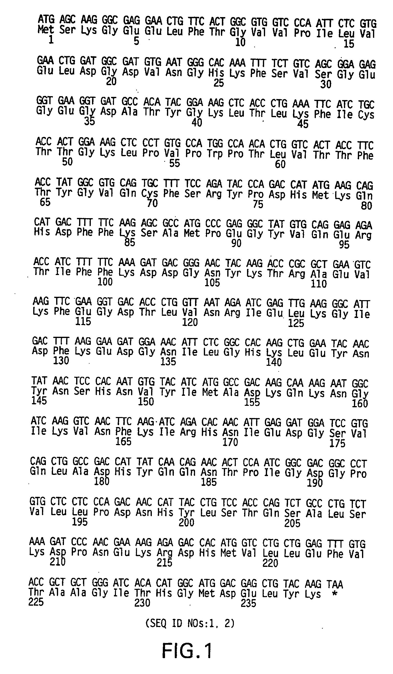
[0080] Plasmids. As depicted in FIG. 5, pGreenLantern-1 (Invitrogen Corporation, Carlsbad, Calif.; catalogue no. 10642) contains the humanized S65T mutant GFP cDNA (FIG. 1; SEQ ID NOs:1, 2) (Evans, K., et al., FOCUS 18(2):40-43 (1996); Zolotukhin, S., et al., J. Virol. 70:4646-4654 (1996)). This plasmid serves as the source of the GFP DNA sequence used for mutagenesis. As depicted in FIG. 6, pGreenLantern-2 contains a universal sequencing primer downstream of the CMV promoter along with an NsiI site immediately upstream of the CMV promoter allowing excision of the promoter region. It also contains XbaI, XhoI and HindIII sites in place of the 3′ NotI site in pGreenLantern-1. A stop codon in the 5′ multiple cloning site of pGreenLantern-1 was shifted out of frame to allow possible fusions to GFP in pGreenLantern-2.

[0081] Mutations to GFP cDNA by UDG cloning. PCR was performed in an MJ Research DNA Engine™ thermal cycler using the following conditions:...

example 2

Growth and Transfection of Host Cells With Mutant GFPs

[0083] Cell Culture. Chinese hamster ovary cells (CHO-K1, obtained from American Type Culture Collection (ATCC), Rockville, Md.) were cultured in D-MEM (4,500 milligrams / liter D-glucose with L-glutamine and phenol red) plus 10% fetal bovine serum (FBS), 0.1 millimolar nonessential amino acids, 2.5 units per milliliter penicillin and 2.5 micrograms per milliliter streptomycin (Freshney, R. I., Culture of Animal Cells: A Manual of Basic Techniques, 3rd Ed., New York: Wiley-Liss (1994)). Cells were grown at 37° C. in a 5% CO2 / air incubator. All media and reagents were from Invitrogen Corporation, Carlsbad, Calif.

[0084] Transfection. CHO-K1 cells were plated at 2×10⁵ cells per well into six-well (35 millimeter diameter) plates one day prior to transfection. Immediately before transfection, cells were rinsed with medium containing no serum or antibiotics. LIPOFECTAMINEreagent was diluted into 100 microliters of OPTI-MEM-I Reduced ...

example 3

Characterization of GFP Mutants Expressed in Eukaryotic Cells

[0086] Formalin Fixation. Transfected host cells were rinsed in Dulbecco's Phosphate Buffered Saline (PBS), then fixed in a solution of 10% formalin in PBS for one hour. Formalin was then removed, and cells were rinsed and stored in PBS at 4° C. until being analyzed.

[0087] Fluorescence Microscopy. Formalin-fixed cells were examined and photographed using an inverted phase contrast fluorescence microscope equipped with FITC filters (excitation 475 nm / dichroic 485 nm / barrier 490 nm) and a 50 watt mercury arc bulb at 1.25 volts. A 40×-power adjustable non-phase objective was used for all micrographs, which were taken through blue, neutral and FITC filters using Kodak Ektachrome ASA 400 Daylight (for slides) or Kodak Gold ASA 400 Daylight (for prints). All exposures were for 12 seconds to allow unbiased comparison of fluorescence intensity.

[0088] Flow Cytofluorimetry. Flow cytofluorimetry was performed on transfected CHO-K1...


PUM

PropertyMeasurementUnit
Volumeaaaaaaaaaa
Volumeaaaaaaaaaa
Volumeaaaaaaaaaa

Abstract

The present invention provides mutants of the Green Fluorescent Protein (GFP) of Aequorea victoria. Specifically provided by the present invention are nucleic acid molecules encoding mutant GFPs, the mutant GFPs encoded by these nucleic acid molecules, vectors and host cells comprising these nucleic acid molecules, and kits comprising one or more of the above as components. The invention also provides methods for producing these mutant GFPs. The fluorescence of these mutants is observable using fluorescein optics, making the mutant proteins of the present invention available for use in techniques such as fluorescence microscopy and flow cytometry using standard FITC filter sets. In addition, certain of these mutant proteins fluoresce when illuminated by white light, particularly when expressed at high levels in prokaryotic or eukaryotic host cells or when present in solution or in purified form at high concentrations. The mutant GFP sequences and peptides of the present invention are useful in the detection of transfection, in fluorescent labeling of proteins, in construction of fusion proteins allowing examination of intracellular protein expression, biochemistry and trafficking, and in other applications requiring the use of reporter genes.

Description

CROSS REFERENCE TO RELATED APPLICATIONS [0001] This application is a continuation of U.S. patent application Ser. No. 10 / 668,168, filed Sep. 24, 2003, which is a continuation of U.S. patent application Ser. No. 09 / 472,065, filed Dec. 23, 1999, now U.S. Pat. No. 6,638,732, which is a continuation of U.S. patent application Ser. No. 08 / 970,762, filed Nov. 14, 1997 (now abandoned), which claims the benefit of U.S. Provisional Application No. 60 / 030,935, filed Nov. 15, 1996, the disclosures of which applications are entirely incorporated herein by reference.BACKGROUND OF THE INVENTION [0002] 1. Field of the Invention [0003] This invention is in the fields of molecular and cellular biology. More particularly, the invention is directed to mutants of the genes encoding Green Fluorescent Protein (GFP) and the proteins encoded by these mutants. The mutant GFPs are used to allow detection of eukaryotic and prokaryotic cells transfected or transformed with extrinsic genes, and to label protein...

Claims


Application Information

Patent Timeline
IPC IPC(8): C07H21/04C07K14/435C12N1/21
CPCC07K14/43595
Inventor EVANS, KRISTA
Owner INVITROGEN
Learn More