Method of culturing proliferative hepatocytes
A proliferative, primary hepatocyte technology, applied in the field of cell culture, 3D hepatocyte culture
- Summary
- Abstract
- Description
- Claims
- Application Information
AI Technical Summary
Problems solved by technology
Method used
Image
Examples
Embodiment 1
[0357] Materials and Method
[0358] material
[0359] cell
[0360] Human liver samples were obtained from patients due to primary hepatocellular carcinoma or liver metastasis due to primary hepatocellular carcinoma or liver metastasis due to primary hepatocellular carcinoma or liver metastasis due to primary hepatocellular carcinoma or liver transfer. The research program was carried out under the French legal guidelines and the local institutional Ethics Committee. The clinical features of human liver samples are detailed in Table 1 below.
[0361] Table 1: Clinical features from their liver samples
[0362]
[0363] Human hepatocytes were separated by two-step collagenase perfusion procedure, and substantive cells were maintained in improved William E medium (referred to as WE HH (human liver cells)), which contain penicillin (100u), streptomycin. (100 μg / ml), insulin (15 μg / mL), glutamine (2 mM), albumin (0.1% (w / v)), transferronin (5.5 μg / mL), sodium selenate (5μ...
Embodiment 2
[0428] Materials and Method
[0429] material
[0430] cell
[0431] Constitutional hepatocytes (PHH) were obtained as described above.
[0432] method
[0433] Primary human hepatocyte culture
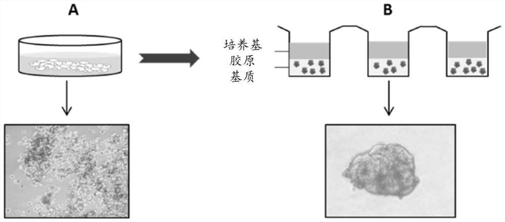
[0434] The primary human hepatocytes 3d cultures were established as described above (see Example 1). In short, PHH was first cultured in a low attachment plate. Then, the aggregate of the obtained pHH is then embedded in the collagen matrix and is further cultured in the collagen matrix.
[0435] Medium
[0436] PHH was cultured in the WE HH medium defined above (see Example 1).
[0437] immunochemistry
[0438] Immunohistochemical staining is performed as described above to evaluate the proliferation of PHH.
[0439] result
[0440] Further studies the importance of growth factor stimulation for the proliferation of PHH in accordance with the 3D culture of the present invention. The pHH in the cultured collagen matrix as described above is deprived of EGF, HGF, and / or ITS (insul...
Embodiment 3
[0453] Materials and Method
[0454] material
[0455] cell
[0456] Sente people hepatocytes (PHH) were obtained as described above (see Example 1).
[0457] method
[0458] Primary human hepatocyte culture
[0459] The primary human hepatocytes 2D and 3D cultures in the collagen matrix are established as described above (see Example 1). PHH was cultured in WE HH medium as described above.
[0460] Comet Determination (Comet Assay)
[0461] The PHH (3D) of (3D) (3d) (3d) (3d) (see Table 7) is incubated with indication concentration (see Table 7) in the control 2D culture (2d) or in a collagen matrix. . After 9 days, PHH was extracted from the collagen matrix by the effect of purified collagenase. The cell precipitate was resuspended in 0.5% low melting agarose and paved on a conventional microscope slide covered with conventional agarose. The electrophoresis migration treatment was transferred and at least 100 images were obtained using a fluorescent microscope after dyeing...
PUM
| Property | Measurement | Unit |
|---|---|---|
| diameter | aaaaa | aaaaa |
| concentration | aaaaa | aaaaa |
| strength | aaaaa | aaaaa |
Abstract
Description
Claims
Application Information
 - R&D
- Intellectual Property
- Life Sciences
- Materials
- Tech Scout
- Unparalleled Data Quality
- Higher Quality Content
- 60% Fewer Hallucinations
Browse by: Latest US Patents, China's latest patents, Technical Efficacy Thesaurus, Application Domain, Technology Topic, Popular Technical Reports.
© 2025 PatSnap. All rights reserved.Legal|Privacy policy|Modern Slavery Act Transparency Statement|Sitemap|About US| Contact US: help@patsnap.com


