
Medicine for treating spinal cord injury, medicine kit and method

A spinal cord injury and kit technology, applied in the field of biomedical technology, can solve problems such as limited functional recovery, and achieve the effects of promoting functional recovery, improving regenerative ability, and enhancing regenerative ability

Pending Publication Date: 2020-07-10
NANTONG UNIVERSITY
View PDF7 Cites 2 Cited by
  • Summary
  • Abstract
  • Description
  • Claims
  • Application Information

AI Technical Summary

Problems solved by technology

Relying on the above two pathways, many scholars have achieved axon regeneration across long-distance injured areas, but only coupling the above two pathways, the functional recovery brought about is still limited

Method used


Image

  • Medicine for treating spinal cord injury, medicine kit and method
  • Medicine for treating spinal cord injury, medicine kit and method
  • Medicine for treating spinal cord injury, medicine kit and method

Examples


Embodiment 1

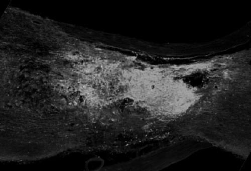
[0031] Example 1. Construction of pinch injury model with overexpression of OPN, IGF1 and T10 in neurons

[0032] Healthy adult female SD rats, SPF grade, weighing 220-250 g. After the rats were weighed, compound anesthetic (0.3ml / 100g) was injected intraperitoneally. After the rats were completely anesthetized, their backs were shaved and disinfected with povidone iodine. The skin and muscle were incised in sequence, and the T8 segment of the spinal cord was located through the bony landmarks. Part of the lamina was removed to expose the spinal cord. The upper and lower vertebrae were fixed through rat spinal cord adapters. OPN and AAV-IGF1 were injected into the spinal cord parenchyma through a digital three-dimensional injection device. A total of 8 injection points were evenly distributed along both sides of the T8 segment. Each injection point was injected at 3 depths, respectively 1.5mm, 1.0mm, and 0.5mm (Virus titer: 1.15×10 13 gc / ml, injection volume: 200nl / point,...

Embodiment 2

[0033] Example 2. Preparation and transplantation of embryonic spinal cord neural stem cells (NSCs)

[0034] 14 days pregnant green fluorescent rats were executed by decapitation after deep anesthesia. After the ventral side was disinfected with 75% alcohol, the skin and muscles were cut open in turn to fully expose them. The fetuses were taken out and placed in a petri dish containing pre-cooled PBS. Identify GFP fetal mice with fluorescent detection lamps; cut off the head and tail of the identified fetal mice, transfer them to a clean petri dish, place the ventral side down, stretch the limbs, find the dorsal midline under a dissecting microscope, and microscissor Gently cut the transparent skin layer from the head to the tail to expose the spinal cord, completely separate the spinal cord from the surrounding tissues, cut off, place in a clean petri dish, and wash 2-3 times in pre-cooled saline; Under the microscope, the dura mater, blood vessels, and possibly attached DR...

Embodiment 3

[0036] Example 3, small molecule compound CLP290 intervention

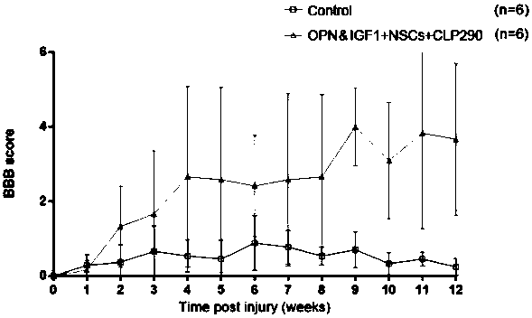
[0037] From the 4th week after operation, 0.2ml small molecular compound CLP290 (concentration: 7mg / ml) was injected intraperitoneally every day until the end of the experiment.


PUM


Abstract

The invention discloses application of growth promoting factor (OPN and IGF1) overexpression in combination with embryo spinal nerve stem cell (NSCs) transplantation and a small molecule compound (CLP290) in preparation of a medicine for treating spinal cord injury. A medicine kit for treating spinal cord injury comprises AAV-OPN, AAV-IGF1, NSCs and CLP290. The invention also discloses applicationof OPN and IGF1 overexpression in combination with NSCs transplantation and the small molecule CLP290 in treatment of spinal cord injury. OPN and IGF1 overexpression, NSCs transplantation and small molecule CLP290 intervention are combined for treating spinal cord injury, the intrinsic regeneration capacity of neurons is enhanced, meanwhile, interrupted upstream and downstream neural loops are rebuilt, the excitability of inhibitory neurons is adjusted, and functional recovery after spinal cord injury is promoted.

Description

technical field [0001] The invention belongs to the field of biomedical technology, and relates to medicines, medicine kits and methods for treating spinal cord injuries, in particular to the application of overexpressed OPN, IGF1 combined with transplanted NSCs and small molecule CLP290 in the preparation of medicines for treating spinal cord injuries, and a method for treating spinal cord injuries. Spinal cord injury drug kit and the application of overexpression of OPN, IGF1 combined with transplantation of NSCs and small molecule CLP290 in the treatment of spinal cord injury. Background technique [0002] With the continuous development of the socio-economic level, the incidence of spinal cord injury is increasing year by year. Spinal cord injury often leads to severe dysfunction of the limbs below the injured segment, causing great pain to the patient, and at the same time, the time and money invested in medical care also bring a heavy burden to the patient's family. A...

Claims


Application Information

Patent Timeline
Patent Type & Authority Applications(China)
IPC IPC(8): A61K38/30A61K48/00A61P25/00A61P25/28A61K31/501A61K35/545A61K38/19
CPCA61K31/501A61K38/30A61K38/19A61K35/545A61P25/00A61P25/28A61K48/0008A61K2300/00
Inventor 于彬王亚先巫荣华单琪顾晓松
Owner NANTONG UNIVERSITY
Learn More