
Thromboelastography heparin quantitative detection kit and preparation method thereof

A thromboelastography and quantitative detection technology, which is applied in the field of medical devices, can solve the problems of inability to detect whole blood, quantitative detection of heparin, and inapplicability of bedside detection, so as to achieve convenience for doctors and patients and simplify the inspection operation process

Active Publication Date: 2018-12-11
上海原科实业发展有限公司
View PDF8 Cites 7 Cited by
  • Summary
  • Abstract
  • Description
  • Claims
  • Application Information

AI Technical Summary

Problems solved by technology

[0026] To sum up, in the prior art: when using APTT or PT detection, only when all the proteins in the whole coagulation cascade are intact can the heparin level be accurately measured, but reagents and instruments will affect its sensitivity to heparin, Causes up to four-fold variability in final results between laboratories; protamine sulfate neutralization test only for unfractionated heparin (UFH) monitoring; coagulation activation assay kit (coagulation method) and heparinase-coated reagents The cup cannot quantitatively detect heparin; in addition, APTT or PT and protamine sulfate neutralization test and anti-Xa activity detection and one-step detection are only suitable for detecting the amount of heparin in plasma. When testing, patient samples need to be processed to obtain plasma It cannot be tested directly on whole blood, so it is not suitable for bedside testing

Method used


Image

  • Thromboelastography heparin quantitative detection kit and preparation method thereof
  • Thromboelastography heparin quantitative detection kit and preparation method thereof
  • Thromboelastography heparin quantitative detection kit and preparation method thereof

Examples


Embodiment 1

[0045] S11) Weigh 0.596g of buffer 4-hydroxyethylpiperazineethanesulfonic acid (English abbreviation: HEPES) into a 250ml volumetric flask, dissolve with an appropriate amount of distilled water, add an appropriate amount of NaOH solution with a concentration of 0.1mol / L to carry out Adjust the pH value, add water to the mark, mix well, and prepare a HEPES buffer with a concentration of 10 mmol / L and a pH value of about 7.9;

[0046] S12) draw distilled water to reconstitute the coagulation factor Xa freeze-dried powder, and make the coagulation factor Xa stock solution with a concentration of 1 g / L;

[0047] S13) draw distilled water to reconstitute the coagulation factor activator RVV-V freeze-dried powder to prepare a coagulation factor activator RVV-V stock solution with a concentration of 1 g / L;

[0048] S14) take by weighing the rabbit brain curd fat and add the weight of the rabbit brain curd fat: volume=1:1 HEPES buffer, grind to milky and no obvious particles are obse...

Embodiment 2

[0054] S21) Weigh 1.489g of buffer 4-hydroxyethylpiperazineethanesulfonic acid and put it into a 250ml volumetric flask, dissolve with an appropriate amount of distilled water, add a 0.1mol / L NaOH solution to adjust the pH value, and add water to the mark, mix well, and prepare a HEPES buffer with a concentration of 25 mmol / L and a pH value of about 7.4;

[0055] S22), S23), S24) are respectively the same as steps S12), S13), S14) of Embodiment 1;

[0056] S25) weigh 0.877g of NaCl, 0.4g of glycine, 2.0g of bovine serum albumin, and 2.5g of trehalose, add them to a 100ml volumetric flask, and add an appropriate amount of HEPES buffer to prepare a support solution;

[0057] S26) draw 2.0ml of coagulation factor Xa stock solution, 0.6ml of coagulation factor activator stock solution, and 0.5ml of rabbit brain coagulation lipid stock solution and add it into the volumetric flask containing the support solution;

[0058] S27) Pipet 50 μl of biological preservative Proclin300 and ...

Embodiment 3

[0061] S31) Weigh 2.979g of buffer 4-hydroxyethylpiperazineethanesulfonic acid (English abbreviation: HEPES) into a 250ml volumetric flask, add an appropriate amount of distilled water to dissolve, add an appropriate amount of NaOH solution with a concentration of 0.1mol / L to carry out Adjust the pH value, add water to the mark, mix well, and prepare a HEPES buffer with a concentration of 50 mmol / L and a pH value of about 7.1;

[0062] S32), S33), and S34) are respectively the same as steps S12), S13), and S14) of Embodiment 1;

[0063] S35) Weigh 1.695g of NaCl, 0.6g of glycine, 3.0g of bovine serum albumin, and 3.8g of trehalose, add them into a 100ml volumetric flask, and add an appropriate amount of HEPES buffer to prepare a support solution;

[0064] S36) draw 3.0ml of coagulation factor Xa stock solution, 1.0ml of coagulation factor activator stock solution, and 1.0ml of rabbit brain coagulation lipid stock solution and add it into the volumetric flask containing the sup...


PUM


Abstract

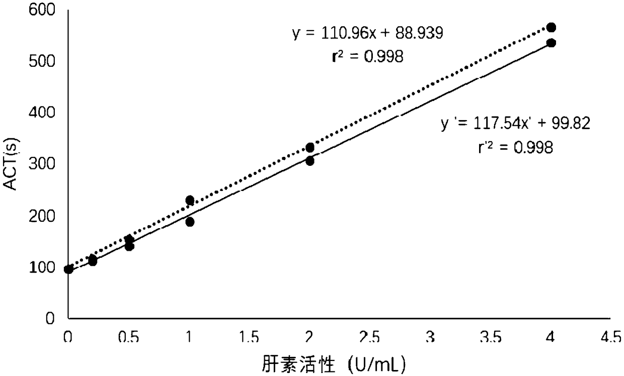
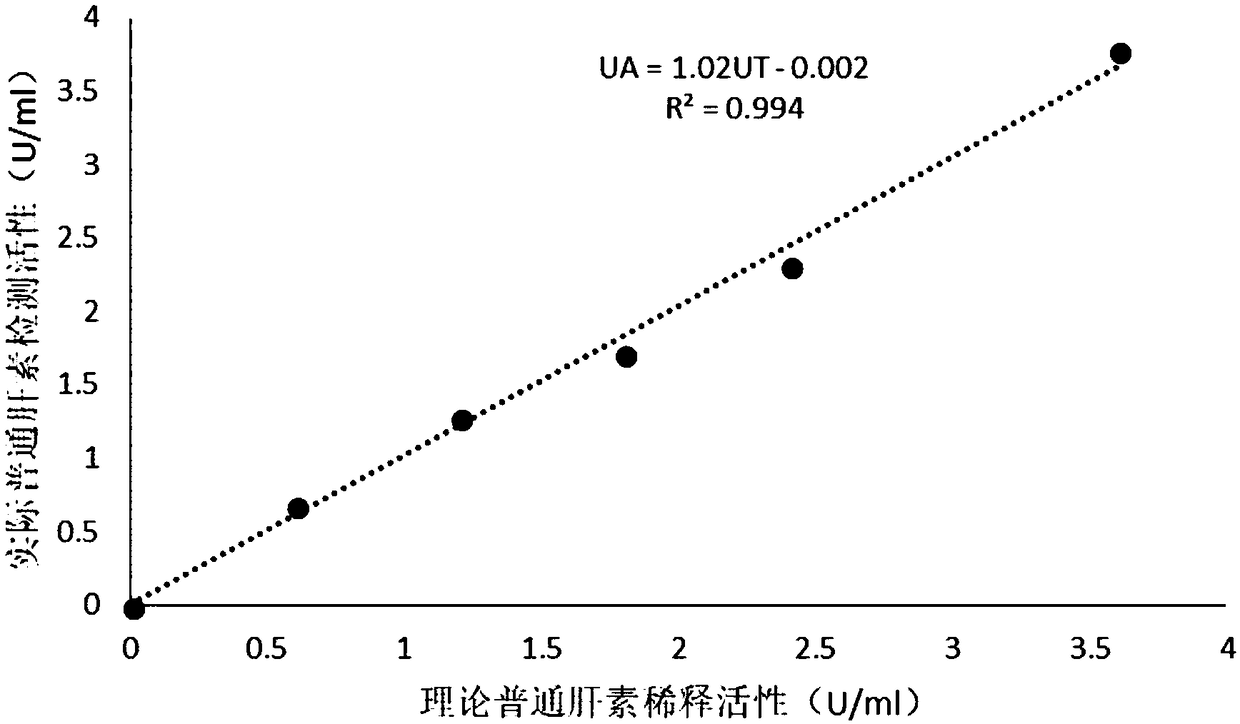
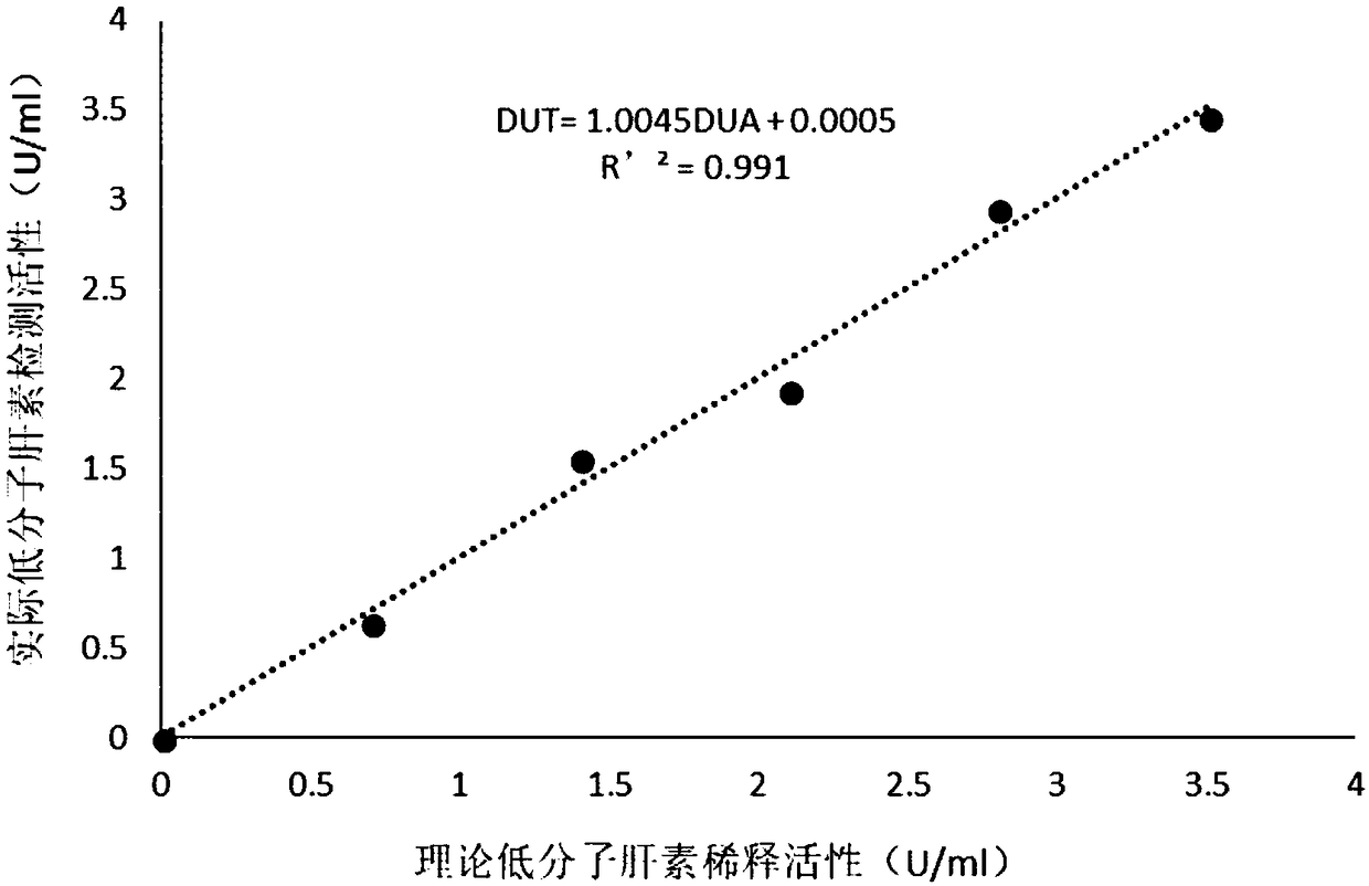
The invention relates to a thromboelastography heparin quantitative detection kit, which comprises a buffer agent, a blood coagulation factor Xa, rabbit brain congealed fat, a coagulation factor activator, a supporting agent, and a biological preservative. The preparation method comprises the following steps: preparing a buffer solution, a blood coagulation factor Xa stock solution, a blood coagulation factor activator stock solution, a biological preservative solution, and a rabbit brain congealed fat stock solution; taking the support agent and adding the buffer solution to prepare a supportsolution; adding the blood coagulation factor Xa, the blood coagulation factor activator, and the rabbit brain congealed fat stock solution to the supporting solution according to the requirements ofkit specification, adding the biological preservative solution after uniformly mixing, then adding the buffer solution to the specified amount, subpackaging and lyophilizing after uniformly mixing. The thromboelastography heparin quantitative detection kit provided by the invention can quantitatively detect the heparin in a sample by a thromboelastography, has good correlation in a linear range,meets the heparin detection limit standard and has a coefficient of variation of not more than 10%, and the sample adopts whole blood without extra treatment; the integrity of protein in a clotting cascade is also not required, and the inspection operating process is simplified, thereby facilitating doctors and patients.

Description

technical field [0001] The invention relates to a thromboelastography heparin quantitative detection kit and a preparation method thereof, belonging to the technical field of medical devices. Background technique [0002] Heparin, named after it was first discovered in the liver, also exists in the lungs, blood vessel walls, intestinal mucosa and other tissues, and is an alternate form of glucosamine, L-iduroside, N-acetylglucosamine and D-glucuronic acid The mucopolysaccharide sulfate composed of it is strongly acidic and is a natural anticoagulant substance in animals. It naturally exists in mast cells and is mainly extracted from bovine lung or pig small intestinal mucosa. [0003] The heparin initially used clinically is called standard heparin, unfractionated heparin or unfractionated heparin (Unfractionated Heparin), with an average molecular weight of 15KD. Antithrombotic, and anticoagulant treatment in hemodialysis, extracorporeal circulation, catheterization, micro...

Claims


Application Information

Patent Timeline
IPC IPC(8): G01N33/66
CPCG01N33/66G01N2400/40
Inventor 王连升李红梅
Owner 上海原科实业发展有限公司
Learn More