Fusion protein for promoting endothelial repair and preparation method and application thereof
A technology of fusion protein and endothelium, applied in the field of genetic engineering, can solve the problems of lack of specificity of implants, delayed endothelialization, etc., and achieve the effects of increasing endothelial inflammation, accelerating endothelialization, and simple preparation method.
- Summary
- Abstract
- Description
- Claims
- Application Information
AI Technical Summary
Problems solved by technology
Method used
Image

Examples
Embodiment 1
[0049] Construction of embodiment 1 fusion protein expression plasmid
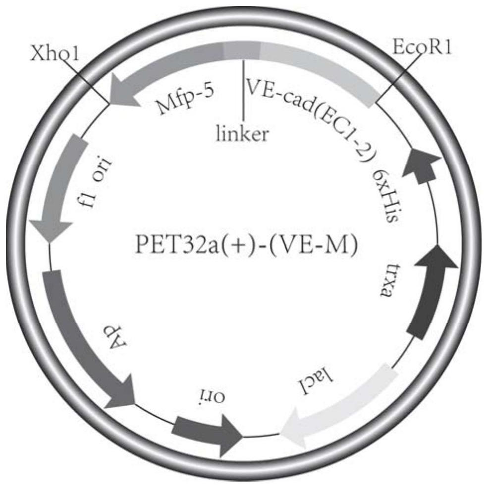
[0050] The present invention utilizes Trizol (Takara Company) to extract total RNA from human umbilical vein endothelial cells (HUVECs), and then uses a reverse transcription kit to reverse the RNA into cDNA. The desired VE-cadherin (EC1-2) fragment is obtained through PCR reaction amplification with specific primers. Due to the difficult source of Mfp-5, the desired Mfp-5 fragment was obtained by chemical synthesis. 15 homologous bases were added at the junction of VE-cadherin (EC1-2) and Mfp-5 sequences, so that VE-cadherin (EC1-2) and Mfp-5 sequences were fused together by In-fusion recombinase, Then cloned into the vector PET32a by T4 ligase, see figure 1 Schematic diagram of fusion protein expression plasmid.
Embodiment 2
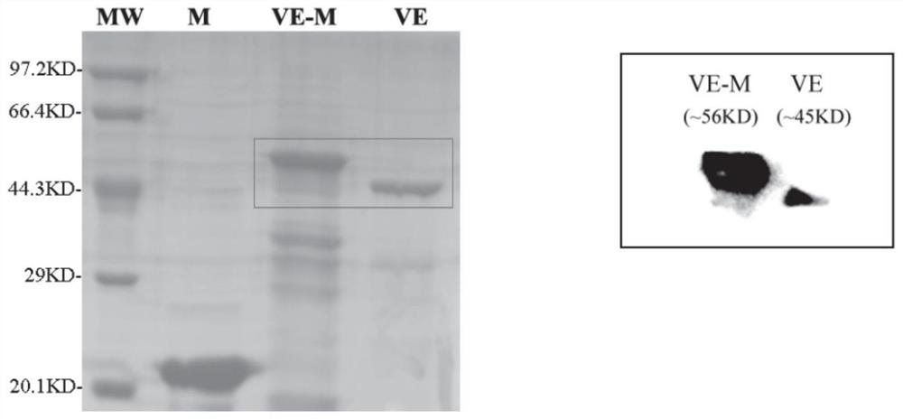
[0051] Expression and purification of embodiment 2 fusion protein
[0052] After the construction of the expression plasmid is completed, the plasmid is introduced into Escherichia coli DE3 (BL21) cells, and according to the amount of required protein, an appropriate amount of Escherichia coli cells containing the target gene expression vector is cultivated to an OD600 of 0.6-0.8, and 0.05mM IPTG is added Induce the expression of the fusion protein at 28°C for 5-8 hours, collect the bacteria by centrifugation at 8000rpm for 20min at 4°C, then sonicate the E. coli cells in an ice bath with a sonicator, collect the supernatant by centrifugation, filter at 0.45um, and pass through His with a nickel column Tags were used to extract and purify the fusion protein, and the resulting protein was dialyzed on ice and stored in 1% HAc. The concentration of the fusion protein was determined by the BCA method and identified by immunoblotting. For the purification of the fusion protein, see...
Embodiment 3
[0053] Embodiment 3 Adhesion experiment of fusion protein and substrate
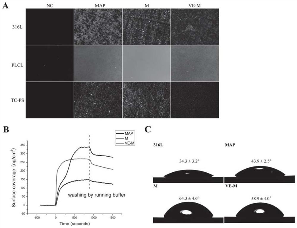
[0054] The present invention locates the fusion protein by combining FITC fluorescent molecules, and measures the adhesion and coating conditions of the fusion protein on three different substrates of 316L stainless steel, PLCL, and TC-PS. In this test, the fusion protein solution is added to Place in 24-well plates with different substrates, incubate at room temperature for 24 hours, take pictures with a fluorescence microscope after ultrasonic cleaning, and characterize the fusion protein coating with an atomic force microscope and a water contact angle meter. At the same time, the adhesion of the fusion protein on the gold sheet was observed in real time by the SPR instrument.
[0055] After soaking, all three kinds of substrates have obvious and uniform fluorescent coatings, which shows that the fusion protein constructed and expressed by the present invention has good adhesion ability to solid subst...
PUM
 Abstract
Description
Claims
Application Information
 - R&D
- Intellectual Property
- Life Sciences
- Materials
- Tech Scout
- Unparalleled Data Quality
- Higher Quality Content
- 60% Fewer Hallucinations
Browse by: Latest US Patents, China's latest patents, Technical Efficacy Thesaurus, Application Domain, Technology Topic, Popular Technical Reports.
© 2025 PatSnap. All rights reserved.Legal|Privacy policy|Modern Slavery Act Transparency Statement|Sitemap|About US| Contact US: help@patsnap.com