Liver capsule wire automatic extracting method based on liver ultrasonic image

An ultrasound image, automatic extraction technology, applied in image enhancement, image analysis, image data processing and other directions, can solve problems such as limited value, misdiagnosis of subjective factors, and missed the best time for treatment

Inactive Publication Date: 2018-06-12
北京华想联合科技有限公司
View PDF1 Cites 4 Cited by
  • Summary
  • Abstract
  • Description
  • Claims
  • Application Information

AI Technical Summary

Problems solved by technology

[0009] Compared with ultrasonography, other imaging examination methods have more or less disadvantages: traditional X-ray imaging lacks sufficient contrast resolution for the evaluation of liver cirrhosis, and its value is limited;
Subjective factors in diagnosis can easily lead to misdiagnosis, or miss the best time for treatment, which may seriously affect the patient's condition and life safety

Method used


Image

  • Liver capsule wire automatic extracting method based on liver ultrasonic image
  • Liver capsule wire automatic extracting method based on liver ultrasonic image
  • Liver capsule wire automatic extracting method based on liver ultrasonic image

Examples


Embodiment 1

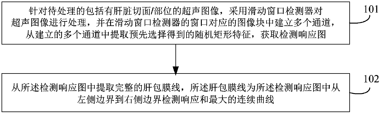
[0101] Such as figure 1 As shown, the method for automatically extracting the liver capsule line based on the liver ultrasound image in this embodiment may include the following steps:

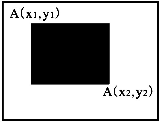
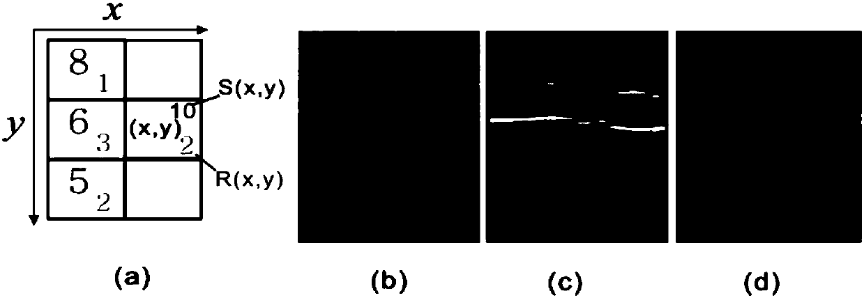
[0102] Step 101, for the ultrasound image to be processed including liver sections / parts, use a sliding window detector to process the ultrasound image, and establish multiple channels in the image block corresponding to the window of the sliding window detector, from the established multiple Extract the pre-selected random rectangular features from channels to obtain the detection response map.

[0103] In this embodiment, the random rectangle features are determined in advance through training samples.

[0104] The ultrasound image in this embodiment may be a pre-acquired test ultrasound image / test image, or a grayscale image obtained in other ways.

[0105] It should be noted that the liver capsule line is extracted in this embodiment, and the above-mentioned ultrasound images are mainly ...

Embodiment 2

[0176] Such as Figure 4 As shown, the method for automatically extracting the liver capsule line of the present embodiment includes the following steps:

[0177] Step 401 , for all training ultrasound images, manually mark the envelope line, uniformly sample and extract image blocks on the envelope line as positive samples, and randomly sample and extract image blocks at other positions of the image as negative samples. Extract a variety of features for positive and negative samples, combine the features and reduce the dimensionality, and train the support vector machine.

[0178] Wherein, the various features are Histogram of Gradients (HOG), Local Binary Pattern (LBP) and Deep Convolutional Neural Network (CNN) features. The structure of CNN is as Figure 5 shown. Figure 5 It is the processing process of the convolutional neural network, and the numbers in the figure are the numbers used in the processing of the convolutional neural network, which are not limited in thi...


PUM


Abstract

The invention provides a liver capsule wire automatic extracting method based on a liver ultrasonic image. The method includes the steps of S1, processing a to-be-processed ultrasonic image containinga liver section / part through a sliding window detector, establishing a plurality of channels in an image block corresponding to a window, and extracting pre-selected random rectangular features to obtain a detection response map; S2, extracting a complete liver capsule wire from the detection response map, wherein the liver capsule wire is a maximum detection response continuous curve from a leftside boundary and a right side boundary in the detection response map. By means of the method, the liver capsule wire can be automatically extracted from the liver ultrasonic image without manual intervention, and furthermore, the accuracy of the capsule wire generated through the method is greatly improved compared with the accuracy of a traditional method.

Description

technical field [0001] The invention relates to image analysis technology, in particular to an automatic liver capsule line extraction method based on liver ultrasound images. Background technique [0002] The liver is an organ in the body that mainly has metabolic functions, and it plays the roles of deoxidation, storage of glycogen, and synthesis of secreted proteins in the body. [0003] Liver cirrhosis is a common clinical chronic progressive liver disease, which is caused by diffuse liver damage caused by one or more pathogenic factors such as viral hepatitis, chronic alcoholism, malnutrition, and intestinal infection for a long time or repeatedly , can be complicated by splenomegaly, ascites, edema, jaundice, esophageal varices, hemorrhage, hepatic coma, can develop into liver cancer, and has a high mortality rate. [0004] Timely detection and treatment with drugs can delay the progress of liver cirrhosis, reduce the incidence of liver cancer, improve the long-term s...

Claims


Application Information

Patent Timeline
IPC IPC(8): G06T7/13
CPCG06T7/13G06T2207/10132G06T2207/20081G06T2207/30056
Inventor 刘翔
Owner 北京华想联合科技有限公司
Why Eureka
Try Eureka