
RGD-peptide-serum protein combined segmental conjugate and preparation method thereof as well as radionuclide marker and application thereof

A radionuclide and serum protein technology, applied in the field of RGD peptide-serum protein binding fragment conjugates, can solve the problems of affecting tumor treatment effect and high side effects

Active Publication Date: 2017-12-15
INST OF NUCLEAR PHYSICS & CHEM CHINA ACADEMY OF
View PDF4 Cites 0 Cited by
  • Summary
  • Abstract
  • Description
  • Claims
  • Application Information

AI Technical Summary

Problems solved by technology

However, due to the lack of radioactive probe drugs that can accumulate and stay at the tumor site for a long time, 177 Lu caused extremely high side effects on normal tissues and metabolic organs during treatment, which seriously affected the efficacy of tumor treatment

Method used


Image

  • RGD-peptide-serum protein combined segmental conjugate and preparation method thereof as well as radionuclide marker and application thereof
  • RGD-peptide-serum protein combined segmental conjugate and preparation method thereof as well as radionuclide marker and application thereof
  • RGD-peptide-serum protein combined segmental conjugate and preparation method thereof as well as radionuclide marker and application thereof

Examples


Embodiment 1

[0058] PEG 4 - Preparation of c(RGDyK): using c(RGDyK) and PEG 4 To connect, the specific synthesis steps are as follows:

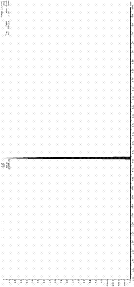
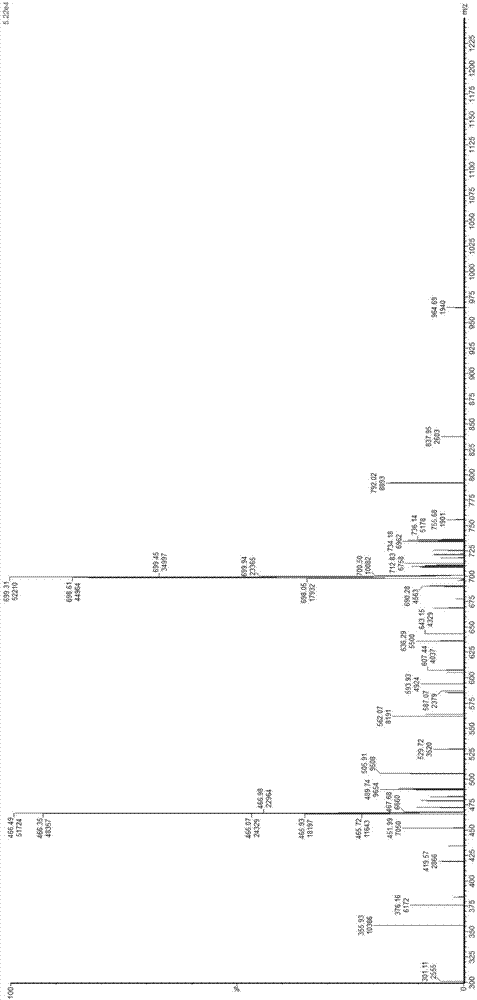
[0059] (1) c(RGDyK) 0.10mmol, PEG 4 0.20mmol, 2-(7-benzotriazole oxide)-N,N,N',N'-tetramethyluronium hexafluorophosphate (HATU) 0.13mmol and diisopropylethylamine 0.50mmol were dissolved in 400 μL of N,N-dimethylformamide (DMF), stirred at room temperature for 1 h, and the resulting reaction solution was separated and purified by preparative high-performance liquid phase (Pre-HPLC).

[0060] (2) Add 3 mL of acetonitrile / water (volume ratio 1:1) to the above reaction solution to obtain a mixed solution, and filter the mixed solution through a 0.22 μm needle filter to obtain a clear solution.

[0061] (3) Separating and purifying the target compound using preparative high-performance liquid chromatography (Pre-HPLC).

[0062] Liquid phase parameters: preparative HPLC column (XBridge BEH C18, 21×250 mm, particle size 5 μm). Solvent A is ultrapure water...

Embodiment 2

[0104] with radionuclides 131 I, Mark III (prepared in Example 1):

[0105] (1) 100 μg / mL aqueous solution of preparation III; Na 131 1 is mixed with the solution of 100mCi / mL with physiological saline;

[0106] (2) 100μL 131 I and 100 μL III were added to a centrifuge tube coated with 1,3,4,6-tetrachloro-3α,6α-diphenylglycoluril (Iodogen), and reacted on a shaker to obtain 131 I-III radionuclide crude product solution.

[0107] (3) Purify the reaction product by HPLC, and the purification parameters are as follows:

[0108] HPLC purification parameters: HPLC column (XDB C18, 4.6×250 mm, particle size 5 μm). Solvent A is ultrapure water containing 0.1% trifluoroacetic acid, solvent B is acetonitrile containing 0.1% trifluoroacetic acid, and the mobile phase gradient is as follows:

[0109]

Flow rate mL / min

A%

B%

0min

1.0

95

5

4min

1.0

95

5

30min

1.0

42

58

35min

1.0

10

90

[0110] UV ...

Embodiment 3

[0120] 131 In vitro stability test of I-III

[0121] 131 After the synthesis of I–III, take 0.5mL (about 1.3mCi) of the purified solution, add it to 3mL of fetal bovine serum, incubate at 37°C for 2h, 8h, 24h, 48h, 72h, and 120h, then perform ITLC analysis. The pure test results are as follows:

[0122] time / h

[0123] Test result: within 72 hours, 131 I-III have good stability. Its radiochemical purity was maintained above 95% as detected by TLC. Its radiochemical purity was maintained above 90% by 8h detection.


PUM

PropertyMeasurementUnit
particle diameteraaaaaaaaaa

Abstract

The invention provides a RGD-peptide-serum protein combined segmental conjugate and a preparation method thereof. The preparation method comprises the following steps: modifying part of tail end of a c(RGDyKRGDyK) lysine chain with a water soluble chain linker (a compound corresponding to R1) to obtain a structural R1-c(RGDyK); modifying c(RGDyK) with a serum protein combined segment, namely, connecting the R1-c(RGDyK) with the serum protein combined segment by using a tri-functional linker (a compound corresponding to R2) to obtain a novel integrin receptor target structure which is named as the RGD-peptide-serum protein combined segmental conjugate. The serum protein combined segment can be combined with serum protein in blood, so that the speed that the modified drug structure by kidney is decreased, and the half life of the drug in vivo is prolonged. The RGD-peptide-serum protein combined segmental conjugate has the structural formula as follows: a formula is as shown in the description.

Description

technical field [0001] The invention belongs to the field of drugs for treating tumors, and in particular relates to an RGD peptide-serum protein binding fragment conjugate combined with integrin and an application thereof. Background technique [0002] Integrin alpha Ⅴ beta 3 It is highly expressed in tumor neovascular endothelial cells, and expressed in many tumors (such as melanoma, glioma, breast cancer, lung cancer), but not expressed in normal tissues and mature vascular endothelium, so it becomes an important Tumor probe binding target. RGD (Arg-Gly-Asp) can interact with integrin α Ⅴ beta 3 It specifically binds, exhibits good permeability, low toxicity, low immune response, and is easy to chemically modify. It is currently the most widely used integrin receptor radioactive imaging polypeptide fragment. Because the molecular structure of RGD and its cyclic derivatives (c(RGDfK) and c(RGDyK)) are too small, they will be rapidly metabolized by the kidneys as radio...

Claims


Application Information

Patent Timeline
Patent Type & Authority Applications(China)
IPC IPC(8): C07K19/00C07K1/06A61K47/64A61K51/08A61P35/00
CPCA61K51/082C07K5/0817C07K14/765C07K2319/00
Inventor 卓连刚赵鹏杨夏廖伟王静王关全魏洪源杨宇川
Owner INST OF NUCLEAR PHYSICS & CHEM CHINA ACADEMY OF
Learn More