
Preparation method of master cell bank for umbilical cord derived mesenchymal stem cells in perinatal period

A master cell bank and mesenchymal stem cell technology, which is applied in the field of preparation of perinatal umbilical cord-derived mesenchymal stem cell master cell bank, can solve the problem of inability to meet the demand for matching bank cell volume, low yield of primary cells, and inability to meet the needs of cells. Quantity requirements and other issues

Inactive Publication Date: 2017-10-10
北京再生生物科技研究院有限公司
View PDF1 Cites 4 Cited by
  • Summary
  • Abstract
  • Description
  • Claims
  • Application Information

AI Technical Summary

Problems solved by technology

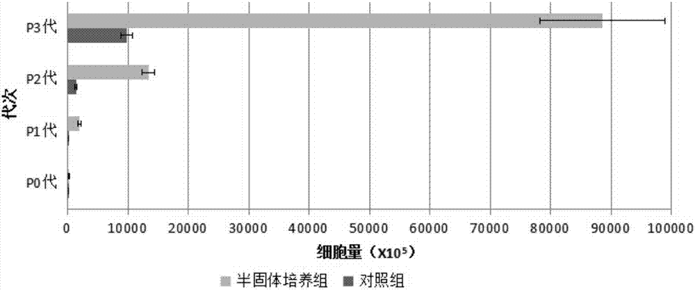
According to the intravenous infusion of P3 generation cell dose 1X10 8 / course of treatment, about 10 courses of treatment, can not meet the "stem cell preparation quality control and preclinical research guidelines (trial)" internal quality control and quality review cell quantity requirements, so the current MSCs bank is still an individualized treatment bank, Unable to meet the cell volume requirements for the preparation of the master cell bank and the working cell bank from the matching bank. The main reason is that the primary cell yield is low due to the backward primary culture process.

Method used


Image

  • Preparation method of master cell bank for umbilical cord derived mesenchymal stem cells in perinatal period
  • Preparation method of master cell bank for umbilical cord derived mesenchymal stem cells in perinatal period

Examples


Embodiment 1

[0056] Example 1: Isolation and culture of MSCs

[0057] S1. Aseptically collect the umbilical cord of a healthy newborn born by full-term cesarean section, the length of the collection is 20cm, both ends are ligated, and the parturient woman signs the informed consent.

[0058] S2. The parturient has been screened for pathogenic microorganisms and has no HIV, HBV, HCV, HTLV, EBV, CMV, or TP infection. The umbilical cord sample was soaked in normal saline, the residual blood sample was collected for sterility test, the result was negative, and the mycoplasma test was carried out, the result was negative.

[0059] S3. Wash the residual blood of the umbilical cord in normal saline, soak it in 75% disinfectant alcohol for about 30 seconds, take it out immediately, rinse it twice with normal saline, cut it off at the ligation site, and discard both ends.

[0060] S4. Perfuse the blood vessel with normal saline, squeeze out the residual blood, cut the umbilical cord sample into ti...

Embodiment 2

[0066] Example 2: Analysis of biological characteristics of P3 generation MSCs

[0067] 2.1 Growth curve and population doubling time detection

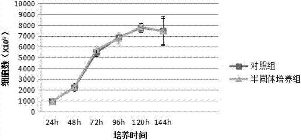
[0068] Take the P2 generation cells, inoculate them into 24-well plates at 1000 / mL, and count 5 wells every 24 hours with a cell imaging system (Cytell CellImaging System, produced by GE Healthcare) for 6 consecutive days. Time is the abscissa, and the growth curve is drawn; the population doubling time is calculated with the formula TD=t×lg2 / lg(Nt / N0) (Nt is the number of cells at time t, and N0 is the number of initial cells).

[0069] 2.2 Cloning rate detection

[0070] Take the P2 cells, inoculate them into 24-well plates at 100 / mL, change the medium in half every other day, culture until the 12th day, discard the culture supernatant, perform HE staining, and count the number of CFU-F.

[0071] 2.3 Detection of differentiation potential in vitro

[0072] 2.3.1 Osteogenic differentiation function

[0073] Take the P2 generatio...

Embodiment 3

[0080] Embodiment three: detection result

[0081] 3.1 Results of yield of umbilical cord-derived MSCs

[0082] On the third day of culture in the 10cm umbilical cord semi-solid culture group, a small amount of cells could be seen crawling out under the microscope, and a large number of cells crawled out on the seventh day of culture, and it was close to 90% confluence on the 12th day of culture. The cell yield after harvesting (X10 5 ) was: 271.91±53.62; on the 12th day of culture in the 10cm umbilical cord control group, an average of 24.2 tissue pieces adhered to the wall, and cells crawled out around them. When harvested on the 14th day, it accounted for about 60% of the culture area, and the cell yield after harvest (X10 5 ) was: 28.91±4.81, significantly less than the semi-solid culture group.

[0083] Take 7.5X10 5 P0 cells at 6000 cells / cm 2 Inoculate to 25cm 2 The culture flask was cultured until harvested on the 4th day, which was recorded as P1 generation cells;...


PUM

PropertyMeasurementUnit
lengthaaaaaaaaaa

Abstract

The invention discloses a preparation method of a master cell bank for umbilical cord derived mesenchymal stem cells in a perinatal period. The preparation method comprises the following steps: S1, collecting a newborn umbilical cord in a sterile way; S2, carrying out pathogenic microorganism screening on a lying-in woman, and carrying out sterile detection on an umbilical cord sample; S3, cleaning the umbilical cord sample, sterilizing, and rinsing with normal saline; S4, carrying out infusion on a blood vessel with the normal saline, squeezing out residual blood, cutting up the umbilical cord sample into tissue blocks, and rinsing; S5, dissolving gelatin with deionized water, carrying out moist heat sterilization, cooling, adding 2XDMEM(L), and uniformly mixing; S6, taking the tissue blocks, adding gelatin solution, transferring into a tissue culture flask, staying overnight in a refrigerator at the temperature of 4 DEG C, adding a complete medium, and putting in a CO2 incubator until cells grow all over a gel layer; and S7, harvesting cells by virtue of a trypsin digestion process, filtering by virtue of a cell strainer, and centrifuging to obtain PO generation MSCs, and cryopreserving the PO generation MSCs, namely an umbilical cord derived MSCs master cell bank is prepared.

Description

technical field [0001] The invention relates to a preparation method of a cell bank, in particular to a preparation method of a perinatal umbilical cord-derived mesenchymal stem cell master cell bank. Background technique [0002] Mesenchymal stem cells (MSCs) are a type of adult stem cells with self-renewal and multilineage differentiation potential, and are one of the most important engineering seed cells, which can be cultured in large scale in vitro without losing self-renewal and multilineage differentiation Potential, it has broad application prospects in drug development, regenerative medicine, tissue engineering, genetic engineering and other fields. [0003] In 2012, PROCHYMAL, developed by Osiris for the treatment of acute graft-versus-host disease in children, was approved for marketing in Canada and New Zealand, which became a landmark event in the history of stem cell regenerative medicine. Subsequently, Hearticellgram-AMI, Cupistem, Cartistem, Osteocel, Trinit...

Claims


Application Information

Patent Timeline
Patent Type & Authority Applications(China)
IPC IPC(8): C12N5/0775
CPCC12N5/0668C12N2509/00
Inventor 张洪钿苑春慧
Owner 北京再生生物科技研究院有限公司
Learn More