
Culture method for gamma delta T cell

A culture method and cell technology, which is applied in the medical field, can solve the problems of insufficient cell expansion, low killing activity, and low expansion, and achieve the effect of increasing cell expansion and increasing killing activity

Inactive Publication Date: 2017-02-15
GUANGZHOU SALIAI STEMCELL SCI & TECH CO LTD
View PDF5 Cites 7 Cited by
  • Summary
  • Abstract
  • Description
  • Claims
  • Application Information

AI Technical Summary

Problems solved by technology

However, the amplification factor of this commonly used method is low, and the killing activity is not high at the same time
[0005] Existing patents CN102994448A and CN103756962A both disclose a method for in vitro expansion of γδT cells, the difference is that the induction medium is different, the former is mononuclear cells containing zoledronic acid, HSP70, IL-7, IL-15, IL -2 cultured in serum-free medium for 12-16 days, the latter is mononuclear cells in the absence of zoledronic acid, HSP70, TLR7 ligand, levamisole, IL-7, IL-15, IL-2 Cultured in serum medium for 12-16 days, although the γδT cells cultured by these two patents have obvious advantages in terms of killing activity, they are not enough in terms of cell expansion multiples, the highest is less than 200 times

Method used


Image

  • Culture method for gamma delta T cell
  • Culture method for gamma delta T cell
  • Culture method for gamma delta T cell

Examples


Embodiment 1

[0028] Example 1: Extraction and separation of peripheral blood mononuclear cells

[0029] Collect 20 mL of human peripheral blood, collect the plasma after centrifugation; dilute the blood cells in the lower layer with normal saline, and then add the diluted solution to the sepmate centrifuge tube (purchased from STEM CELL Company) that already has Ficoll separation solution (purchased from STEM CELL Company), Centrifuge for 10-20min; after centrifugation, pour out the supernatant liquid from the centrifuge tube, then add a certain amount of RPMI 1640 medium to resuspend the cells, and centrifuge to obtain mononuclear cells.

Embodiment 2

[0030] Embodiment 2: cultivation method of the present invention

[0031] Follow 1-2 x 10 6 / mL cell density was added to X-VIVO 15 medium for culture, and IL-15, OKT-3 and IL-2 were added to the medium; on the 6th day, K562 tumor cell lysate was added to stimulate γδT cells; On day 9, add IFN-γ to sensitize γδT cells; 14 days later, obtain γδT cells; pay attention to supplementing medium and cytokines during this period.

[0032] Among them, the concentrations of IL-15, OKT-3 and IL-2 can be selected as follows:

[0033] (1) IL-15 at 500 U / mL, OKT-3 at 100 μg / mL and IL-2 at 500 U / mL;

[0034] (2) IL-15 at 100 U / mL, OKT-3 at 500 μg / mL and IL-2 at 1000 U / mL;

[0035] (3) IL-15 at 1000 U / mL, OKT-3 at 50 μg / mL and IL-2 at 100 U / mL;

Embodiment 3

[0036] Example 3: Detection of the surface marker CD56 of γδT cells by flow cytometry + and CD3-

[0037] Control method: according to 1×10 6 Add X-VIVO 15 medium for culture at a cell density of / mL, and add IL-15 and IL-2 to the medium. After 14 days of culture, γδT cells are obtained; pay attention to supplementing medium and cytokines during this period.

[0038] The inventive method: adopt the method for the first culture medium in embodiment 2;

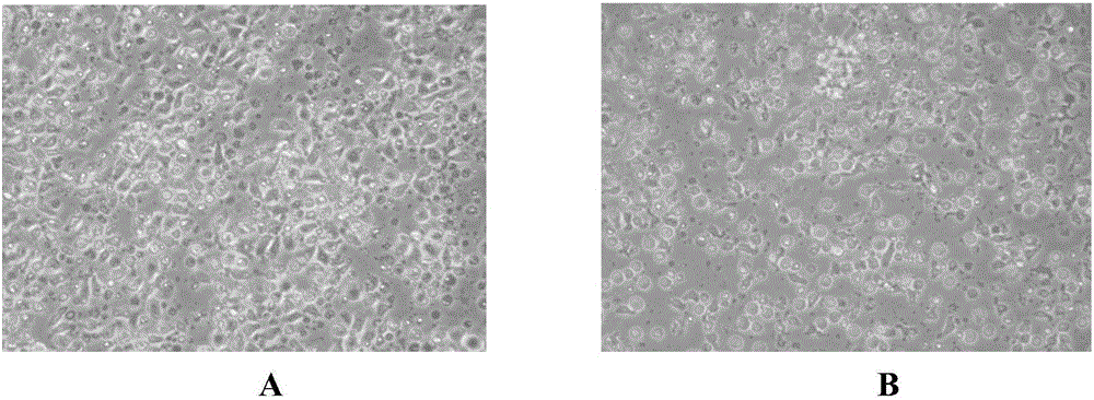
[0039] The γδT cells cultured by the two methods were used for flow cytometric detection of CD56 and CD3. The flow detection method is as follows: take 1×10 6 γδT cells; centrifuge at 250g for 5min to remove the supernatant; wash twice with PBS solution containing 10% FBS; add 2.5μL of CD56 and CD3 antibodies in the dark and incubate at room temperature for 30min; wash twice with PBS solution containing 10%FBS; The cells were resuspended in RPMI 1640 medium and filtered, and tested by flow cytometry, see the results figure ...


PUM


Abstract

The invention relates to the medical field and discloses a culture method for a gamma delta T cell. The culture method comprises the steps of carrying out induced culture on a mononuclear cell containing IL-15, OKT-3 and IL-2 in a serum-free culture medium until the gamma delta T cell is induced, then adding a tumor cell lysis solution, continuing to carry out culture and multiplication, then adding TNF-alpha, continuing to carry out culture to promote the maturity and further multiplication of gamma delta T cell, and culturing for 14 days to obtain the gamma delta T cell. Cell factors of an induced culture medium are regulated in the earlier stage, and stimulation and sensitization are carried out by virtue of the tumor cell lysis solution and TNF-alpha in the middle and later stage of the culture, so that the amplification multiple of the cell is increased, the killing activity is enhanced, and the secretion amounts of IFN-gamma and IL-4 are increased.

Description

technical field [0001] The invention relates to the medical field, in particular to a method for culturing γδT cells. Background technique [0002] Cellular immunity is the main force in the body's process of killing tumors. Cellular immunity is mediated by T lymphocytes, and T lymphocytes are MHC-restricted when killing tumor cells, that is, CD4+T cells can only recognize MHC-II complexes, and CD8+T cells can only recognize MHC-class I complex, two types of T cells cannot recognize cells that do not express the corresponding MHC, so it is limited. Non-restricted tumor killing generally refers to killing tumors without MHC recognition, usually including NK cells and CIK cells. [0003] γδT cells are a type of non-MHC-restricted innate T lymphocytes distributed in peripheral blood and mucosal tissues. They are mainly distributed in intestinal epithelium, skin, spleen and liver, and their proportion in peripheral blood is very low. γδT cells have been proven to be able to e...

Claims


Application Information

Patent Timeline
IPC IPC(8): C12N5/0783
CPCC12N5/0636C12N2501/2302C12N2501/2315C12N2501/24C12N2501/999C12N2502/11C12N2502/30
Inventor 王一飞陈海佳葛啸虎罗二梅王小燕
Owner GUANGZHOU SALIAI STEMCELL SCI & TECH CO LTD
Learn More