
Magnetic resonance imaging method based on magnetization transfer combined with slice-selective inversion recovery pre-pulse

A technology of magnetic resonance imaging and inversion recovery, applied in medical science, sensors, diagnostic recording/measurement, etc., can solve the problems of inability to obtain satisfactory suppression effect, poor background suppression effect, prolonged signal acquisition time, etc., to make up for the inspection. Long time, improved vascular inflow enhancement effect, easy to use effect

Active Publication Date: 2017-10-10
THE FIRST AFFILIATED HOSPITAL OF MEDICAL COLLEGE OF XIAN JIAOTONG UNIV
View PDF4 Cites 0 Cited by
  • Summary
  • Abstract
  • Description
  • Claims
  • Application Information

AI Technical Summary

Problems solved by technology

However, this technology still has many difficulties in MRA of body vessels, which are mainly reflected in the following aspects: ①body vessels run in multiple directions, and small vessels are tortuous, while TOF method is sensitive to the direction of blood flow and is prone to saturation ② The inspection time is too long. For body vascular imaging, in order to avoid breathing and heartbeat artifacts, ensure that the signal acquisition site is at the same position each time, use ECG triggering and breathing triggering or breath-holding if necessary technology; for patients with poor respiratory function or children (cannot hold breath or irregular breathing), it is necessary to increase the acquisition, which will prolong the acquisition signal time; in addition, due to the large imaging range of the body, the acquisition time will also be increased; and Excessive time (10 minutes to 20 minutes) acquisition is intolerable to the subject, and the result is poor imaging effect; ③Tissue with short T 2 * Especially in the lungs, the air-lung tissue interface makes the magnetic field gradient in the lungs uneven, resulting in susceptibility artifacts that affect the observation of lung tissue. Similarly, the signal of small blood vessels in the lung is also very low, which limits the observation; ④Poor background suppression: The conventional TOF method uses a fast gradient echo sequence with short TR and short TE to saturate the background tissue through repeated radiofrequency stimulation to achieve the effect of background suppression. TE is the echo time (time of Echo), Refers to the time interval between a radio frequency pulse and the corresponding echo
However, for short T1 (longitudinal relaxation time) tissues (such as adipose tissue), it can restore the longitudinal magnetization vector in a short time, and cannot obtain satisfactory suppression effect
Therefore, MRA of the body TOF method is not effective in background suppression due to the interference of chest wall and mediastinal fat

Method used


Image

  • Magnetic resonance imaging method based on magnetization transfer combined with slice-selective inversion recovery pre-pulse
  • Magnetic resonance imaging method based on magnetization transfer combined with slice-selective inversion recovery pre-pulse
  • Magnetic resonance imaging method based on magnetization transfer combined with slice-selective inversion recovery pre-pulse

Examples


Embodiment Construction

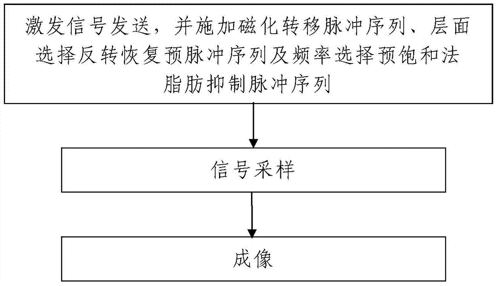
[0043] Such as figure 1 A magnetic resonance imaging method of magnetization transfer combined with layer-selective inversion recovery pre-pulse is shown, which uses a magnetic resonance imaging device to perform non-contrast agent-enhanced angiography under free breathing, and the method includes the following steps:

[0044] Step 1. Sending excitation signals: using the magnetic resonance imaging equipment to send excitation signals to the detected object, and the transmitted excitation signals are scanning imaging sequences.

[0045] When sending the excitation signal, the magnetic resonance imaging equipment is used to apply the magnetization transfer pulse sequence and the slice selective inversion recovery pre-pulse sequence according to the combination of magnetization transfer and layer selective inversion recovery.

[0046] Step 2. Signal sampling: the magnetic resonance imaging equipment is used to sample the magnetic resonance data of the detected object, and the ma...


PUM


Abstract

The invention discloses a magnetic resonance imaging method of magnetization transfer combined with layer selection inversion recovery pre-pulse, comprising steps: 1. Excitation signal transmission: sending an excitation signal to a detected object; when sending an excitation signal, select according to magnetization transfer and layer Combining inversion recovery, applying magnetization transfer pulse sequence, layer-selective inversion recovery pre-pulse sequence and frequency-selective pre-saturation fat suppression pulse sequence; 2. Signal sampling: using magnetic resonance imaging equipment to detect the magnetic resonance imaging of the detected object. The data is sampled, and the sampled magnetic resonance data is the echo signal of the transmitted excitation signal; 3. Imaging: obtaining blood vessel image data according to the sampled magnetic resonance data. The method of the invention has simple steps, reasonable design, convenient implementation, and good application effect. On the premise of not using contrast agent for enhancement, high-quality blood vessel imaging can be obtained and the imaging time is short. Angiography methods.

Description

technical field [0001] The invention relates to a magnetic resonance imaging method, in particular to a magnetic resonance imaging method using magnetization transfer combined with layer selective inversion recovery pre-pulse. Background technique [0002] Blood vessels are the conduits for biological transport of blood, and are an important physiological and anatomical basis for maintaining metabolism in the human body. As we all know, various diseases will lead to morphological changes of blood vessels, and pathological changes of blood vessels can also cause corresponding target organ involvement. Therefore, blood vessels are closely related to the occurrence, development and outcome of various diseases and are mutually causal. How to evaluate and display the physiological and pathological changes of blood vessels in vivo is very important for in-depth investigation of the cause, development, prognosis assessment and treatment plan decision-making of diseases, and has alw...

Claims


Application Information

Patent Timeline
Patent Type & Authority Patents(China)
IPC IPC(8): A61B5/055
Inventor 杨健牛刚李贤军王蓉孙亲利郭建新荐志洁
Owner THE FIRST AFFILIATED HOSPITAL OF MEDICAL COLLEGE OF XIAN JIAOTONG UNIV
Learn More