
Method for establishing monoclonal mesenchymal stem cells and its application

A kind of stem cell and cell technology, applied in the field of biomedicine, can solve the problems of no cell surface labeling, complicated operation, influence of cell activity, etc.

Active Publication Date: 2017-08-25
张文炜
View PDF1 Cites 0 Cited by
  • Summary
  • Abstract
  • Description
  • Claims
  • Application Information

AI Technical Summary

Problems solved by technology

The disadvantages of the first two methods mainly lie in the low purity of cells; the common disadvantages of the latter two methods are that 1) there is no real specific cell surface marker; 2) they have a great impact on cell viability, which can easily cause damage to MSCs and cause proliferation Slowness and other issues; 3) The operation is complicated and expensive, generally limited to laboratory applications
But the disadvantages are: the bone marrow source is inconvenient to obtain materials, and it is easy to cause autologous injury; the peripheral blood source must rely on exogenous stimulation to increase the number of stem cells; although the fat source avoids the above disadvantages, it has the tumorigenicity of in vitro expansion and the use of serum Pathogenicity

Method used


Image

  • Method for establishing monoclonal mesenchymal stem cells and its application
  • Method for establishing monoclonal mesenchymal stem cells and its application
  • Method for establishing monoclonal mesenchymal stem cells and its application

Examples


Embodiment 1

[0035] Example 1 Cell Separation and Culture

[0036] Different tissues derived from animals were digested with collagenase, after centrifugation, and the supernatant was removed, the collected cells were washed twice with PBS and then cultured with complete medium. Change the medium and remove the rest of the suspended cells to obtain all the adherent cells. The complete medium includes α-MEM culture medium, 10% fetal bovine serum.

Embodiment 2

[0037] Example 2 Cell transfection

[0038] The newly isolated MSCs were adhered to the wall and cultured for 24 hours, and continued to culture after changing the medium. Before transfection, they were cultured in an antibiotic-free medium. After the cells were 70%-80% confluent, the plasmid containing the Nanog promoter and the control plasmid were dissolved separately. In 50 microliters of serum-free Opti-MEMI medium, mix well. Dissolve an appropriate amount of liposomes in 50 microliters of serum-free Opti-MEM Ⅰ culture medium, and mix well. Incubate for 5 min at room temperature. The above two solution mixtures were then mixed (total volume 100 µl). Mix gently and incubate at room temperature for 25 minutes, add 100 microliters of the complex and appropriate amount of medium to each well of a 24-well plate. After adding the complex for 4-6 hours, replace the medium with complete medium, and keep the cells at 37 degrees 5% CO 2 After culturing in the incubator for 48 h...

Embodiment 3

[0040] Example 3 Cell Sorting

[0041] After digestion and centrifugation with trypsin, the adherent cells after successful transfection were transferred from the culture dish with PBS, and after washing, they were detected by a FACS flow cytometer under an argon laser at 488 nm. 10,000 cells in the sample were analyzed using Cell-Quest software (Weasel V2.3.1).

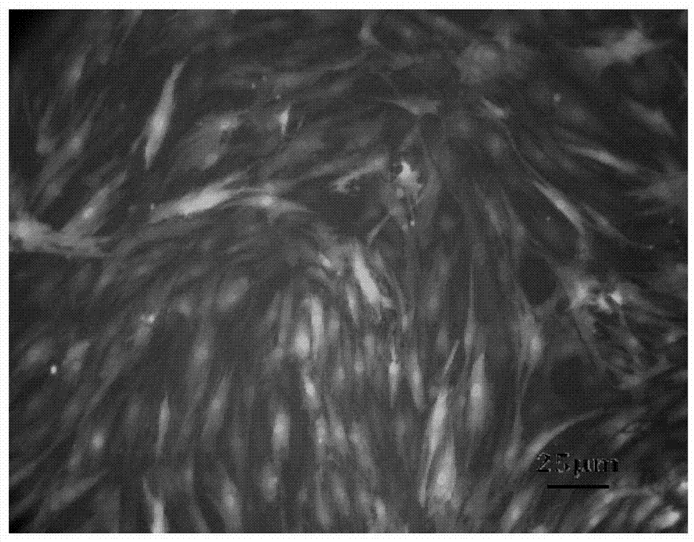
[0042] The sorted single cells were cultured in a 96-well plate at 37°C and 5% CO using the above-mentioned complete medium. 2 Cultivate under the same conditions and change the medium every 3 to 5 days. It can be seen from the observation that the growth of individual cloned cells is relatively slow in the early stage, but after a week, the cells grow rapidly after forming a colony group, showing a typical mesenchymal stem cell-like growth, showing a spindle shape , swirl-like, radial growth, the results are shown in image 3 .


PUM


Abstract

The invention proposes a method for constructing monoclonal mesenchymal stem cells and its application. A method for isolating mesenchymal stem cells, comprising: A) digesting an animal sample with collagenase to disperse cells contained in said animal sample so as to obtain a cell mixture containing stem cells; B) converting a nucleic acid molecule encoding a reporter protein to Introduced into at least a portion of the cells of the mixture of cells, wherein the nucleic acid molecule encoding the reporter protein is operably linked to a mesenchymal stem cell-specific promoter so as to express the reporter protein in the mesenchymal stem cells; and C ) using FACS to sort and obtain the mesenchymal stem cells expressing the reporter protein, wherein the mesenchymal stem cells are in the form of single cells. Using this method, mesenchymal stem cells in the form of single cells can be efficiently prepared.

Description

technical field [0001] The present invention relates to the field of biomedicine. In particular, the present invention relates to methods for isolating stem cells and uses thereof. More specifically, the present invention relates to a method for isolating stem cells, the stem cells or their derivatives obtained by the method, and the use of the stem cells or their derivatives in the preparation of medicines. Background technique [0002] Mesenchymal stem cells (MSCs) have recently received a lot of attention because of their properties that facilitate transplantation, namely their multipotency and non-immunogenicity. It has been reported that mesenchymal stem cells (MSCs) can be isolated from adult bone marrow and fetal tissues. MSCs isolated from bone marrow (BM MSCs) have been reported as pluripotent cells with high proliferative potential in vitro. BMMSCs are capable of differentiating into adipocyte (A), osteoblast (O), chondrocyte (C), and vascular smooth muscle (V) ...

Claims


Application Information

Patent Timeline
Patent Type & Authority Patents(China)
IPC IPC(8): C12N5/0775A61K35/28A61P7/00A61P37/00A61P25/00A61P3/10A61P9/00A61P19/00
CPCA61K35/28A61P3/10A61P7/00A61P9/00A61P19/00A61P25/00A61P37/00C12N5/0662C12N2510/00
Inventor 张文炜
Owner 张文炜
Learn More