
Ultrasonic probe, medical endoscope and processing method for ultrasonic probe and medical endoscope

An ultrasonic probe and endoscope technology, applied in the field of medical endoscopes and ultrasonic probes, can solve the problems of shortening the service life of the probe, poor chemical resistance, corrosion of solder joints, etc. Impact capability, the effect of prolonging the service life

Inactive Publication Date: 2014-02-05
SONOSCAPE MEDICAL CORP
View PDF13 Cites 0 Cited by
  • Summary
  • Abstract
  • Description
  • Claims
  • Application Information

AI Technical Summary

Problems solved by technology

However, the chemical resistance of polyurethane is relatively poor. When the endoscope is frequently soaked, disinfected and cleaned, especially for veterinary endoscopes, the polyurethane insertion tube is prone to cracking and peeling, resulting in the scrapping of the entire endoscope.
Although the chemical medium resistance of silicone rubber material is better than that of polyurethane, due to the longer bond length and larger bond angle of the silicon-oxygen bond, its permeability resistance is not good, especially when the medium molecule is relatively small, it is extremely easy to penetrate silicon. The rubber insertion tube penetrates into the electronic part of the endoscope
In addition, because polyurethane and silicone rubber materials are not smooth enough and have a large friction coefficient, when they enter the human body, it is easy to cause discomfort to the patient and increase the difficulty of the doctor's operation.
[0008] 2. Regarding the cable sheath material of medical equipment, although the commonly used polyvinyl chloride material is cheap and resistant to certain chemical media, its hardness is relatively high and the hand feeling is relatively poor
[0009] 3. Regarding the acoustic lens material of ultrasonic probes and ultrasonic endoscopic probes, silicone rubber with a sound velocity lower than water and better softness is generally used at present. Based on the characteristics of silicone rubber itself and the requirements for the acoustic impedance of acoustic lenses, the general hardness can only reach About 55A, some high-frequency probes are easy to damage the transducer due to the thin lens, even a slight bump
Moreover, acoustic coupling agents and small molecule disinfectants, such as glutaraldehyde, ethanol, isopropanol, etc., easily penetrate into the matching layer through the silicone rubber acoustic lens, destroying the bonding layer between the matching layer and the piezoelectric ceramics and the piezoelectric ceramics and the piezoelectric ceramics. The adhesive layer of the backing material will peel off the matching layer of the probe, the piezoelectric ceramics and the backing material. In severe cases, it will corrode the solder joints of the piezoelectric ceramics, making the image cannot be displayed normally, and greatly shortening the service life of the probe. The main reasons for the current probe damage

Method used


Image

  • Ultrasonic probe, medical endoscope and processing method for ultrasonic probe and medical endoscope
  • Ultrasonic probe, medical endoscope and processing method for ultrasonic probe and medical endoscope
  • Ultrasonic probe, medical endoscope and processing method for ultrasonic probe and medical endoscope

Examples


Embodiment 1

[0045] The embodiment of the present invention provides a durable and not easily damaged ultrasonic probe and medical endoscope.

[0046] like Figure 5 , 6 As shown, they are the schematic structural diagrams of the medical endoscope 1 and the ultrasonic probe 2 respectively. Both the medical endoscope probe and the ultrasonic probe have acoustic lenses 11 and 21 at the front end respectively, and the acoustic lenses 11 and 12 usually have a hardness of about 55A. silicone rubber material. Most of the insertion tube 12 of the endoscope is made of polyurethane. Endoscopes and ultrasound probes generally use PVC cable sheaths with relatively high cost performance13, 22, some manufacturers use polyurethane cable sheaths, and a few high-quality endoscopes and ultrasound probes use silicon. Cable with rubber sheath.

[0047]

[0048] The solution described in the specific embodiment of the present invention is to arrange a layer of protective film on the outer surface of the u...

Embodiment 2

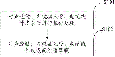
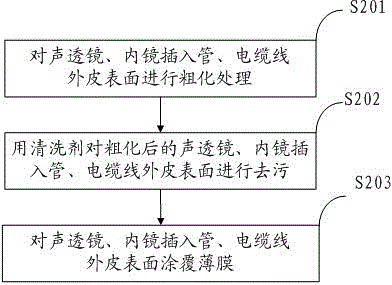
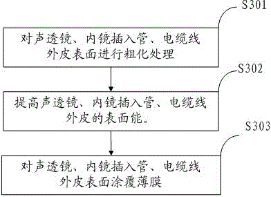
[0057] like figure 1 As shown, the present invention also provides a method (hereinafter referred to as the method) for setting a protective film on the surface of the ultrasonic probe, the acoustic lens of the medical endoscope, the endoscope insertion tube thereof, and the outer skin of the cable (hereinafter referred to as the method), and the method includes the following steps:

[0058] S101. Roughen the surface of the acoustic lens, its endoscope insertion tube, and the outer skin of the cable.

[0059] The specific method of the roughening treatment can use the physical roughening method. In this specific embodiment, the sandblasting method is preferred, and the insertion tube, the cable sheath and the surface of the acoustic lens are roughened, and the angle of the sandblasting is controlled at 25-75°. °, the particle size of the sand is selected at 400-900 mesh. After roughening, use compressed air to blow off the sand, and then repeatedly wipe the surface with deio...


PUM

PropertyMeasurementUnit
Thicknessaaaaaaaaaa
Hardnessaaaaaaaaaa

Abstract

The embodiment of the invention provides an ultrasonic probe and a medical endoscope. Two layers of protective films are respectively arranged on outside surfaces of an acoustical lens 21 and a cable sheath 22 of the ultrasonic probe and outside surfaces of an acoustical lens 11, an endoscope insert tube 12 and a cable sheath 13 of the medical endoscope. Meanwhile, the invention also provides a method for installing the protective films outside the acoustical lenses, the endoscope insert tube and the cable sheaths. By adopting the technical scheme provided by the invention, the ultrasonic probe and the medical endoscope can be more durable and are not easy damage, the ultrasonic probe and the medical endoscope can be helped to be cleaned and disinfected, and the friction between equipment and a human body is reduced.

Description

[0001] technical field [0002] The invention relates to the technical field of medical endoscopes and ultrasonic probes, in particular to an ultrasonic probe, a medical endoscope and a processing method thereof. Background technique [0003] Medical endoscope (hereinafter referred to as medical endoscope) is a tube equipped with light, which can enter the stomach through the mouth or enter the body through other natural orifices. The endoscope can be used to see the lesions that X-rays cannot show, so it Very useful for doctors. Endoscopy usually includes ordinary endoscopy and ultrasonography combined with ultrasound. [0004] An ultrasound probe is a device used to perform ultrasound scans and is used in conjunction with ultrasound equipment for ultrasound imaging. [0005] Currently, if Figure 5 , 6 So, are the structural schematic diagrams of the medical ultrasonic endoscope 1 and the ultrasonic probe 2 respectively. The endoscopic probe and the ultrasonic probe bot...

Claims


Application Information

Patent Timeline
IPC IPC(8): A61B8/00A61B1/00H01B13/22
Inventor 王乐杨佳陈雄
Owner SONOSCAPE MEDICAL CORP
Learn More