Schmallenberg virus nucleocapsid protein monoclonal antibody, and preparation method thereof
A technology of monoclonal antibody and nucleocapsid protein, which is applied in the field of genetic engineering and immunity to achieve the effect of abundant expression, high purity and good repeatability
- Summary
- Abstract
- Description
- Claims
- Application Information
AI Technical Summary
Problems solved by technology
Method used
Image

Examples
preparation example Construction
[0052] 2 Preparation of immune splenocytes
[0053] After 3 days of booster immunization, aseptically collect splenocytes to prepare splenocyte suspension, the specific operation is as follows:
[0054] 1) Mice were killed by dislocation;
[0055]2) Soak the mouse in 75% alcohol for disinfection, cut its abdomen with sterile scissors, take out the spleen, put it in a sterile plate containing a small amount of culture solution (1640 culture solution) and put it on a stainless steel drying net. The needle core was ground into a cell suspension and counted;
[0056] 3) Collect the spleen cell suspension on ice, centrifuge to remove the supernatant;
[0057] 4) Wash the pellet again by centrifugation with fresh RPMI-1640 culture medium;
[0058] 5) Add fresh RPMI-1640 culture medium; count spleen cells.
[0059] 3 Recovery of myeloma cells
[0060] The myeloma cell SP2 / 0 was stored in a liquid nitrogen tank at -196°C, and it was sufficient to recover, proliferate, and passage...
Embodiment 3
[0105] Example 3 Anti-SBV N Protein Monoclonal Antibody Titer Determination——ELISA Detection
[0106] 1. Coating: The purified recombinant N protein was used as the detection antigen, the coating concentration was 2 μg / ml, 100 μL / well, overnight at 4°C, and washed 3 times with the washing solution.
[0107] 2. Blocking: add 150 μL / well blocking solution, wash 3 times after 2 hours at 37°C, and pat dry. Store in a 4°C refrigerator for later use.
[0108] 3. Add the sample to be tested:
[0109] 1) For serum / antibody / ascitic fluid titer detection, dilute the first well at 1:1000, then dilute downward at a gradient ratio of 1:2, incubate at 37°C for 30 minutes, wash the plate 4 times, and pat dry.
[0110]2) For cell supernatant detection, pipette 100 μL of cell supernatant, add it to the corresponding microplate, incubate at 37°C for 30 min, wash the plate 4 times, and pat dry.
[0111] 3) Add secondary antibody: take horseradish-enzyme-labeled goat anti-mouse IgG (IgG-specif...
Embodiment 4
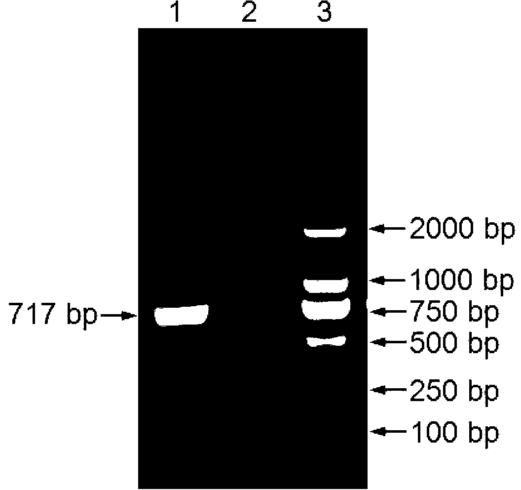
[0117] Example 4 Western blot identification of anti-Schmallenberg virus nucleocapsid protein N monoclonal antibody (2C8)
[0118] 1 Purpose of the test
[0119] To verify whether the monoclonal antibody 2C8 can react with the recombinant N protein and the natural nucleocapsid protein (N) of SBV.
[0120] 2 test material
[0121] Cell line: BHK-21.
[0122] SBV strains: BH80 / 11-4, BH652 / 12-1, BH619 / 12, D512 / 12, D495 / 12-1, and D495 / 12-2; viral stock was 10 6 TCID 50 / mL.
[0123] HRP-goat anti-mouse IgG (product number P0447): purchased from Dako Company in Denmark.
[0124] 3 operation steps
[0125] 3.1 Preparation of protein samples
[0126] Cells infected by different strains of SBV and control cells (approximately 2.0×10 6 cells / bottle) were digested from the wall of the cell flask, transferred to a 15mL centrifuge tube with a pipette, centrifuged at 300×g at 4°C for 6min, poured off the cell supernatant, and resuspended the cell pellet with 2mL PBS. Store in a 4°...
PUM
 Abstract
Description
Claims
Application Information
 - R&D
- Intellectual Property
- Life Sciences
- Materials
- Tech Scout
- Unparalleled Data Quality
- Higher Quality Content
- 60% Fewer Hallucinations
Browse by: Latest US Patents, China's latest patents, Technical Efficacy Thesaurus, Application Domain, Technology Topic, Popular Technical Reports.
© 2025 PatSnap. All rights reserved.Legal|Privacy policy|Modern Slavery Act Transparency Statement|Sitemap|About US| Contact US: help@patsnap.com