


Quick Research
Generate reliable direction feasibility study reports for your R&D in just a few steps.

Technical Q&A
Discover and master advanced knowledge NOW. Basics, ideas, possibilities, all at once.

Find Solutions
As an expert in R&D theories, this can generate solutions to your technical problems instantly.

Evaluate Feasibility
Analyze your overall solution with one click, know your potential R&D risks in advance.

Monitor Landscape
Get weekly tech updates, stay abreast of the latest tech innovations and key insights.
Integration micro-fluidic chip for immune analysis research and applications thereof
A microfluidic chip and immune analysis technology, applied in the field of immune analysis, can solve the problems of long analysis time and cumbersome steps, and achieve the effects of improving analysis efficiency, shortening reaction time, and reducing consumption
- Summary
- Abstract
- Description
- Claims
- Application Information
AI Technical Summary
Problems solved by technology
Method used
Image

Examples
Embodiment 1
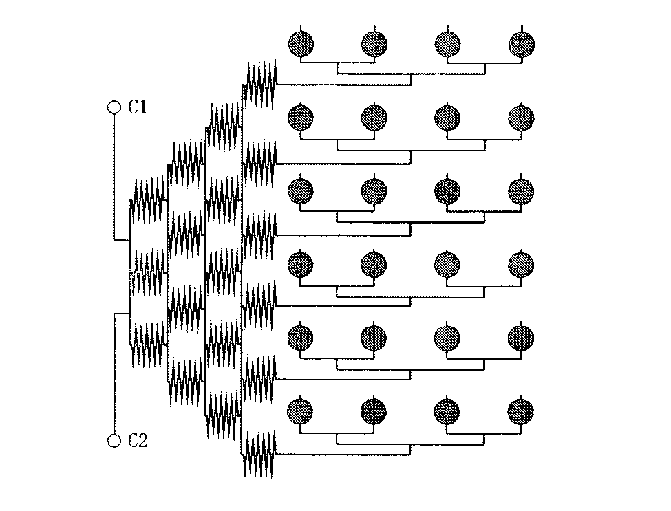
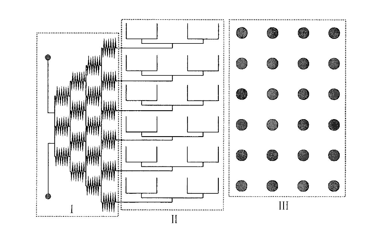
[0028] The microfluidic chip is used for the research application of the optimal concentration of drugs. image 3 After surface modification of the chip shown with BSA-glutaraldehyde-Protein A, 5 μg / The antibody in ml was passed into the PDMS channel and allowed to stand for 60 minutes, and then passed into TBS buffer to wash away the unfixed antibody. Pass 5 μg / ml antigen into the channel and let it stand for 20 minutes, then pass through TBS buffer to wash away unfixed antigen. Pass 5 μg / ml of secondary antibody into the channel and let it stand for 20 minutes, then pass through TBS buffer to wash away unfixed secondary antibody. Pass through 4-MUP with a drug concentration of 0.0001 mmol / l to react for 3 minutes, and then place it in a microplate reader to detect the fluorescence intensity (excitation wavelength and emission wavelength are 365nm and 448nm, respectively). Repeat the above operation, respectively change the concentration of the drug 4-MUP to 0.001, 0.01, 0...
Embodiment 2
[0030] The application of microfluidic chips in the study of drug action time. image 3 After surface modification of the chip shown with BSA-glutaraldehyde-protein A, 5 μg The / ml antibody was passed into the PDMS channel and allowed to stand for 60 minutes, and then passed into TBS buffer to wash away the unfixed antibody. Pass 5 μg / ml antigen into the channel and let it stand for 20 minutes, then pass through TBS buffer to wash away unfixed antigen. Pass 5 μg / ml of secondary antibody into the channel and let it stand for 20 minutes, then pass through TBS buffer to wash away unfixed secondary antibody. After passing through 4-MUP with a drug concentration of 0.10mmol / l, place it under a microplate reader to detect the fluorescence intensity (excitation wavelength and emission wavelength are 365nm and 448nm respectively). The experimental results are as follows: Figure 5 shown.
Embodiment 3
[0032] Application of microfluidic chip in the study of inhibition rate of enzyme inhibitors. image 3 After surface modification of the chip shown with BSA-glutaraldehyde-protein A, 5 μg The / ml antibody was passed into the PDMS channel and allowed to stand for 60 minutes, and then passed into TBS buffer to wash away the unfixed antibody. Pass 5 μg / ml antigen into the channel and let it stand for 20 minutes, then pass through TBS buffer to wash away unfixed antigen. Pass 5 μg / ml of secondary antibody into the channel and let it stand for 20 minutes, then pass through TBS buffer to wash away unfixed secondary antibody. One inlet of the chip is fed with 0.10mM 4-MUP and inhibitor KH 2 PO 4 buffer, and the other inlet into 0.10mM 4-MUP and 1mM KH 2 PO 4 React for 3 minutes, and then place it under a microplate reader to detect the fluorescence intensity (excitation wavelength and emission wavelength are 365nm and 448nm, respectively). Experimental results such as Figure ...
PUM
 Abstract
Description
Claims
Application Information
 - R&D Engineer
- R&D Manager
- IP Professional
- Industry Leading Data Capabilities
- Powerful AI technology
- Patent DNA Extraction
Browse by: Latest US Patents, China's latest patents, Technical Efficacy Thesaurus, Application Domain, Technology Topic, Popular Technical Reports.
© 2024 PatSnap. All rights reserved.Legal|Privacy policy|Modern Slavery Act Transparency Statement|Sitemap|About US| Contact US: help@patsnap.com