
Preparation method of blood vessel support or cardiac valve surface coating with good biocompatibility

A biocompatible, vascular stent technology, applied in the field of biochemical modification, can solve the problems of increased vascular stent, social loss, and poor anticoagulant function on the surface of the stent

Inactive Publication Date: 2009-02-11
SOUTHWEST JIAOTONG UNIV
View PDF0 Cites 11 Cited by
  • Summary
  • Abstract
  • Description
  • Claims
  • Application Information

AI Technical Summary

Problems solved by technology

But there are still some problems: with the prolongation of drug-coated stent implantation, the drug inhibits the proliferation of smooth muscle cells (the main cause of restenosis) and also inhibits the proliferation of endothelial cells, and the surface of the stent cannot form a complete endothelium. The anticoagulant function on the surface of the stent is poor; and the final drug release is exhausted and the complete endothelial cell layer that has the anticoagulant effect and inhibits the proliferation of smooth muscle cells is still not formed, and the possibility of restenosis in the vascular stent thereafter is significantly increased
[0005] At present, there are about 200,000 patients in our country who need artificial heart valve replacement surgery every year due to serious valvular disease. Most of these patients are young and middle-aged. If the valve cannot be replaced in time, it will cause immeasurable losses to the society.

Method used


Image

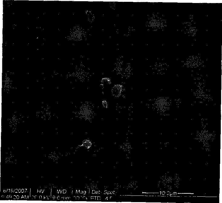
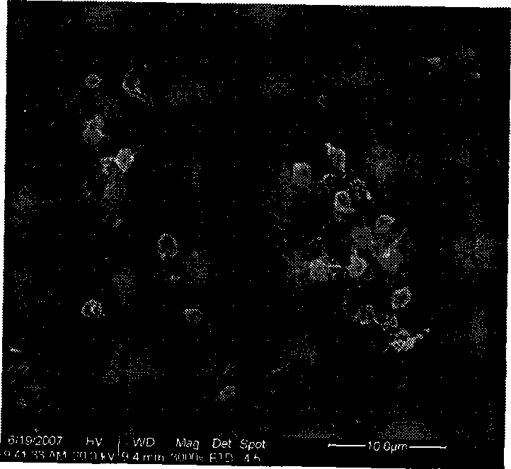
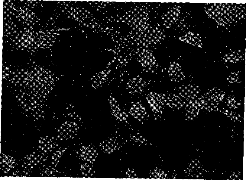
  • Preparation method of blood vessel support or cardiac valve surface coating with good biocompatibility
  • Preparation method of blood vessel support or cardiac valve surface coating with good biocompatibility
  • Preparation method of blood vessel support or cardiac valve surface coating with good biocompatibility

Examples


Embodiment 1

[0024] A preparation method of a vascular stent surface coating with good biocompatibility, the steps are:

[0025] A. Preparation of coating solution Dissolve heparin in deionized water, adjust its pH value to 7, and add endothelial cell growth factor to prepare a heparin / growth factor solution; the heparin concentration in the heparin / growth factor solution in this example 0.5mg / ml, endothelial cell growth factor concentration is 5ng / ml.

[0026] The collagen is dissolved in 0.2 mol / L acetic acid solution to prepare a collagen solution, and the concentration of the collagen solution is 0.5 mg / ml.

[0027] Before performing the dip-coating in the following step B, first immerse the vascular stent in a 0.5mol / L strong alkali solution at 60°C for 3 hours for activation; take it out, ultrasonically clean it with deionized water, and then immerse it in a 0.1mg / ml poly In the polylysine solution, soak for 1 hour, take out the deionized water to clean, blow dry with nitrogen, and ...

Embodiment 2

[0031] A method for preparing a heart valve surface coating with good biocompatibility, the steps are:

[0032] A. Preparation of coating solution Dissolve heparin in deionized water, adjust its pH value to 8, and add endothelial cell growth factor to prepare a heparin / growth factor solution; the heparin concentration in the heparin / growth factor solution is 25 mg / ml, the concentration of endothelial cell growth factor is 50ng / ml.

[0033] The collagen is dissolved in 0.2 mol / L acetic acid solution to prepare a collagen solution, and the concentration of the collagen solution is 5 mg / ml.

[0034]Before the dip coating of the following step B, the heart valve is first activated by immersing it in a 5mol / L strong alkali solution at 80°C for 24 hours; take it out, ultrasonically clean it with deionized water, and then immerse it in 10mg / ml polymer In the lysine solution, soak for 12 hours, take out the deionized water to clean, blow dry with nitrogen, and then carry out the dip...

Embodiment 3

[0038] A preparation method of a vascular stent surface coating with good biocompatibility, the steps are:

[0039] A. Preparation of coating solution Dissolve heparin in deionized water, adjust its pH value to 7.5, and add endothelial cell growth factor to obtain a heparin / growth factor solution; the heparin concentration in the heparin / growth factor solution is 5mg / ml, the concentration of endothelial cell growth factor is 10ng / ml.

[0040] The collagen is dissolved in 0.2 mol / L acetic acid solution to prepare a collagen solution, and the concentration of the collagen solution is 1 mg / ml.

[0041] Before the dip-coating of the following step B, the stent is activated by immersing it in a 3mol / L strong alkali solution at 70°C for 10 hours; take it out, ultrasonically clean it with deionized water, and then immerse it in 2mg / ml polymer In the lysine solution, soak for 4 hours, take out the deionized water to clean, blow dry with nitrogen, and then carry out the dip coating i...


PUM


Abstract

The invention discloses a preparation method of a surface coating of intravascular stents or cardiac valves with excellent biocompatibility, which includes the steps: A. the preparation of a coating solvent: heparin is dissolved in deionized water, the pH value thereof is adjusted to 7 to 8 and then an endothelial cell growth factor is added to form a heparin / growth factor solution; collagens are dissolved in acetum to form a collagen solution; B. dip-coating: the intravascular stents or the cardiac valves are immersed in the heparin / growth factor solution and a layer of heparin / growth factor is dipped and coated on the surface of the intravascular stents or the cardiac valves, and then the intravascular stents or the cardiac valves are taken out for being cleaned by the deionized water and being dried by nitrogen gas; and the intravascular stents or the cardiac valves are immersed in the collagen solution, coated with a layer of collagen, and taken out for being cleaned by the deionized water and being dried by nitrogen gas; the steps of dip-coating by the use of the heparin / growth factor and the collagen are repeated in sequence for 1 time to 60 times; finally the step of dip-coating by the use of the heparin / growth factor is carried out again to obtain the surface coating with excellent biocompatibility of the intravascular stents or the cardiac valves. The intravascular stents or the cardiac valves prepared have excellent biocompatibility and low preparation cost.

Description

Technical field [0001] The invention relates to a method for biochemically modifying the surface of a vascular stent and a heart valve material. Background technique [0002] The incidence of cardiovascular disease is increasing year by year. Atherosclerosis causes stenosis or obstruction of the lumen is the main cause of ischemic heart disease (coronary heart disease); heart valve disease is due to congenital or acquired reasons The resulting heart valve disease causes heart blood flow disorder-based disease. [0003] The latest research proves that endothelial cells are very important for the function of blood vessels: 1) The intact endothelial cell layer will secrete nitric oxide and other molecules to inhibit the excessive proliferation of smooth muscle, and the secretion mechanism of the damaged endothelial cell layer is out of balance, leading to the excessive proliferation of smooth muscle and resulting in vascular 2) The intact endothelial cell layer will secrete an...

Claims


Application Information

Patent Timeline
IPC IPC(8): A61L27/40A61L27/34
Inventor 黄楠陈佳龙李全利王进杨苹冷永祥陈俊英孙鸿万国江赵安莎游天雪吴熹
Owner SOUTHWEST JIAOTONG UNIV
Learn More