
Brachytherapy method of treating skin tumors using a tailor-made radioactive source

a skin tumor and brachytherapy technology, applied in radiation therapy, radiation therapy, x-ray/gamma-ray/particle irradiation therapy, etc., can solve the problems of significant increase in the risk of developing subsequent bcc's at other sites, insufficient information provided by traditional diagnostic methods, and especially vulnerable rims of the ear and the lower lip, so as to prevent any possible skin contamination, and control the effect of irradiation

Active Publication Date: 2016-11-08
ONCOBETA INT GMBH
View PDF13 Cites 3 Cited by
  • Summary
  • Abstract
  • Description
  • Claims
  • Application Information

AI Technical Summary

Benefits of technology

[0048]In one embodiment, the method further comprises the step of calculating, i.e. accurately calculating, the radiation dose distribution curve to be delivered using a dosimetric program or an algorithm.
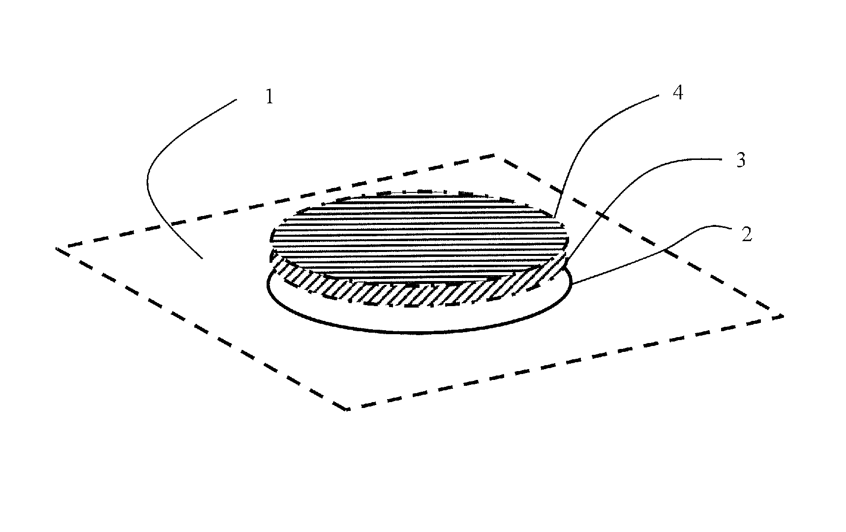
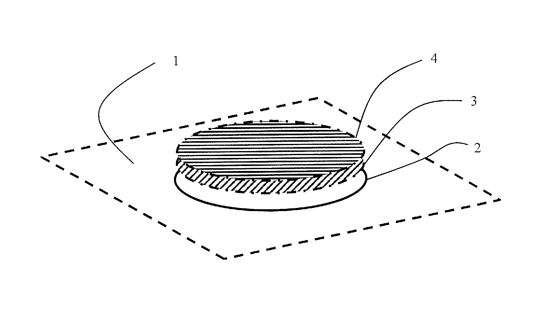
[0056]The “protective film” advantageously is uniform and continuous and easily adheres to the surface of the area of skin to be treated. This film protects the subject from any possible radioactive contamination and from any physical contact with the radioactive source. By such a film, irradiation of the skin lesion can be performed without physically touching the skin of the patient with any radioactive source material.
[0057]The “protective foil” also adheres to the surface of the area of skin to be treated and protects the subject from radioactive contamination and physical contact with the radioactive source. Usually, it is a thin plastic foil which is directly applied to the area of skin to be treated to protect and insulate the skin.
[0059]Here, a medical application of radioactive sources useful for radiotherapy of skin tumors in human and animals is provided. To our knowledge, none prior-art document discloses a medical method, aimed or directed to the therapy of skin tumors by shaping a selective, individually tailor-made brachytherapy source for the irradiation of the cancerous lesions. In the present proposed method, differently from the sources currently used in classical brachytherapy with metallic radioactive plaques or seeds or needles, the obtained sources are moldable, to assume different shapes and thicknesses, and can be safely used without contamination of the physician and the patient. By starting from a solution containing a radioactive isotope, a solid matrix is obtained, in which the radioactive isotope is embedded; the matrix in which the isotope is immobilized is sufficiently flexible and variable to adapt to different surfaces and substrates. The matrix which encloses the radioactive isotope is chosen in order to be chemically compatible with the embedded isotope, and to ensure homogeneity of the imparted dose. The radioactive sources obtained in this way can be safely and effectively used in the therapy of skin tumors, with excellent cure rate and with negligible side effects.
[0061]In this respect, the use of beta radiation isotopes embedded in a tailor-made irradiation mold, like the technique proposed in the present application, can override, in skin tumors, the drawbacks of classical radiotherapy or brachytherapy, which make use for irradiation of gamma or X-rays photons. By using for therapeutic treatments a tailor-made source, like the ones proposed and realized in the present application, not only the margin of irradiation can be easily controlled by the application of the resin only on the lesion area, but the dose distribution curve obtained by beta particles, almost ideally follows the typical distribution of the tumor invasion in the dermal tissue. This fact allows an administration of therapeutic doses only at the required depth, without unnecessary dose deposition in the sub dermal tissue, independently from the lesion shape complexity, and from the number of the lesions. The product can be easily applied on the protected skin, always obtaining a regular and smooth surface of uniform thickness. The protection of the skin by a thin layer of an accurately calculated amount of protective cream, chosen in the group of creams or gels containing one or more hydrophilic components, or by a layer of a thin plastic adherent foil applied to the lesion, was important to prevent any possible skin contamination. Moreover, by using the described method, the material of the radioactive source doesn't in any way touch the skin, and the sources are not classified as radiopharmaceutical products. The total dose imparted from the beta emitting layer depends on the amount of radioactive nanocolloid; the mass unitary dose depends on the ratio (radioactivity of colloid) / (weight of resin); the unitary surface dose depends on the ratio (radioactivity of colloid) / (area of applied resin). The amount of resin necessary to a uniform spreading is approximately 10 microliters / square cm (μl / cm2). The thickness of the resin must be taken into account in the dosimetric program for a precise dose evaluation, by calculating the self-absorption effect.

Problems solved by technology

Once a person has developed a BCC, there is a significantly increased risk of developing subsequent BCC's at other sites.
There are several different histological and pathological clinical forms of BCC, but traditional diagnostic methods do not provide enough information on tumor features.
Both tumors appear most frequently on the face, neck, bald scalp, hands, shoulders, arms and back; the rim of the ear and the lower lip are especially vulnerable to these cancers.
In all cases in which tumors are located in areas on which surgery excision may be very difficult (ear, nose, eyelids), the aesthetic and functional results are often highly unsatisfactory.
The cosmetic outcome is often unsatisfactory; if a relapse raises in transplanted skin, the management of the lesions becomes highly problematic.
Irradiation by conventional methods (radiotherapy by external beam X-rays or gamma rays), due to the penetrating nature of the photons, however, cannot be recommended for treatment of tumors in areas in which radiation can be very harmful (face, eyes), and has proven itself unsatisfactory in the treatment of SCC.

Method used


Image

  • Brachytherapy method of treating skin tumors using a tailor-made radioactive source
  • Brachytherapy method of treating skin tumors using a tailor-made radioactive source

Examples


example 1

Composition and Methods for the Fabrication of Radioactive Sources for Radiotherapy of Skin Tumors

[0064]Among the possible modes for obtaining radioactive sources in thin layer, a useful technique consists in evaporating a volatile solution, containing both a coordination complex of the isotope and a dissolved matrix-forming molecule (i.e. a plastic, or a gum, or a polymer). By this technique, however, a uniformity of dispersion of the isotope during the solvent evaporation is hard to obtain, due to the fact that the evaporation of a solvent is not a regular process. Another technique used consists in the use of a polymerizable matrix (epoxy resin, etc.); in this case the semi-solid form of the matrix renders problematic the fabrication of a regular thin layer of uniform thickness.

[0065]The present application proposes as a matrix for the preparation of radioactive source in thin layer for use in medicine, the use, as a binding matrix, of a semi-fluid resin or paint, preferably chos...

example 2

Application of the Radiotherapy of Skin Tumors

[0071]More than 300 patients with histologically confirmed diagnosis of BCC or of SCC were enrolled for a brachytherapy treatment, by using the radioactive sources prepared by one of the above mentioned methods. In many of the patients a relapse of the tumor successive to a surgical excision was present; in others a surgical operation would have been impossible, or functionally and / or aesthetically unacceptable. The application of the product must be preceded by an accurate cleaning and curettage of the lesions. This treatment is useful not only for more precisely delineating the extent of a tumor, but also to eliminate all the keratinic crusts, granulation tissue, scabs, that, due to their thickness, would stop the beta particles irradiation. Both BCC and SCC appear often ulcerated, with presence of serous and blood scabs; in other cases keratin plaques or nodules of horny consistence are present. In all cases an accurate and complete e...

example 3

Medical Results

[0078]Immediately after the treatment, a faint reddening of the treated skin area is visible. After a few days a variable erythema is present, sometimes with emission of serum, and a crust or scab is formed. An apparent worsening of the aspect of the lesion is often observed, with the appearance of a burn, but the bleeding, often present before the therapy, usually disappears. After 40-120 days the erythema fades, sometimes a second scab is formed, and itch is present. The clinical healing is more clearly apparent, the tumor neoangiogenic development, often clearly visible with epiluminescence before the treatment, starts to disappear. After 60-180 days, in most of the cases, an apparent clinical healing is present, rarely with persistence of a scab. The lesion area becomes paler than the untreated skin, the tumor neoangiogenic development disappears, and a fading in the skin coloration appears.

[0079]A Perspex screen 10 mm thick was found sufficient to protect the phy...


PUM


Abstract

Embodiments of the invention relate to a method of treating a cancerous or non-cancerous skin lesion of a subject, e.g. a human patient, by epidermal radioisotope therapy, a specialized type of brachytherapy. The method can include defining and marking an area of skin to be treated; covering the area with a protective layer, e.g. a protective film or foil; applying a tailor-made radioactive source by applying a layer of a radioactive source material on the protective layer, such that the area is covered by the material while any area not to be treated is spared; and removing the radioactive source after a predetermined time period of irradiation.

Description

CROSS-REFERENCE TO RELATED APPLICATION[0001]The present application claims priority under 35 U.S.C. §119(a) of Australian Patent Application No. 2010200556, filed on Feb. 15, 2010, which is hereby incorporated by reference in its entirety, including any figures, tables, or drawings.FIELD OF THE INVENTION[0002]The present invention refers to a brachytherapy method of treating a tumor skin lesion, preferably a basal cell carcinoma or a squamous cell carcinoma, comprising the step of applying to an area of skin to be treated a tailor-made radioactive source comprising a matrix-forming component, preferably an acrylic resin, and a radioactive isotope, preferably a beta emitter isotope, on a film or foil covering said area in order to protect the skin from radioactive contamination.BACKGROUND OF INVENTION[0003]Skin cancer is the most common form of cancer in humans, and in some countries it accounts for about half of all tumors. Among all tumors of humans, basal cell carcinoma (BCC) is t...

Claims


Application Information

Patent Timeline
Patent Type & Authority Patents(United States)
IPC IPC(8): A61N5/00A61N5/10
CPCA61N5/1028A61N5/1029A61P35/00
Inventor CIPRIANI, CESIDIODESANTIS, MARIA
Owner ONCOBETA INT GMBH
Learn More