RGD-(bacterio)chlorophyll conjugates for photodynamic therapy and Imaging of Necrotic tumors
a technology of rgd chlorophyll and necrotic tumors, applied in the field of oncology, can solve the problems of poor prognosis of cancer patients, tumor aggressiveness, hypoxic and eventually necrotic, and out-growth of oxygen supply in cancer patients
- Summary
- Abstract
- Description
- Claims
- Application Information
AI Technical Summary
Benefits of technology
Problems solved by technology
Method used
Image
Examples
example 1
Transfection of Tumor Cells with Fluorescence Proteins
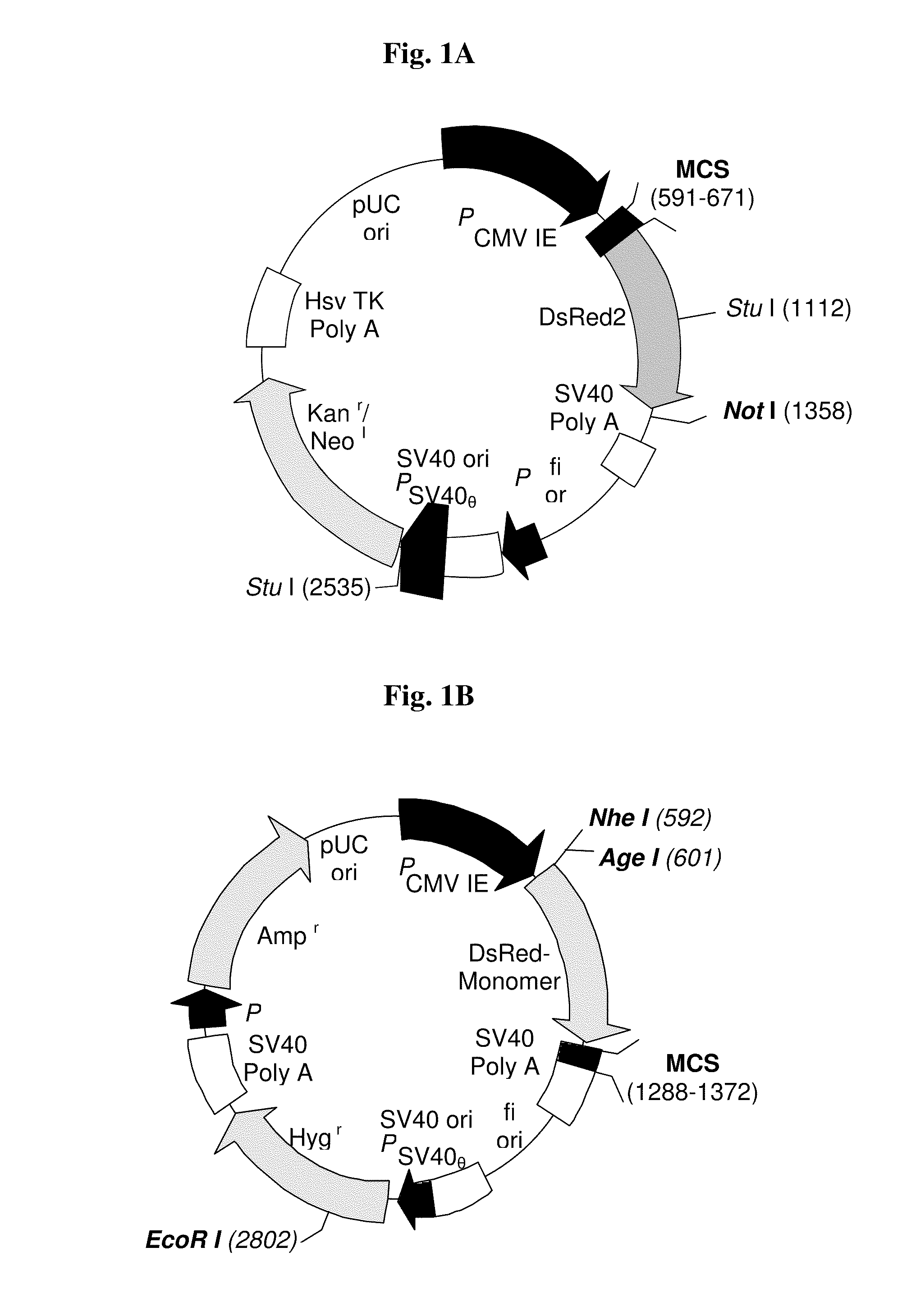
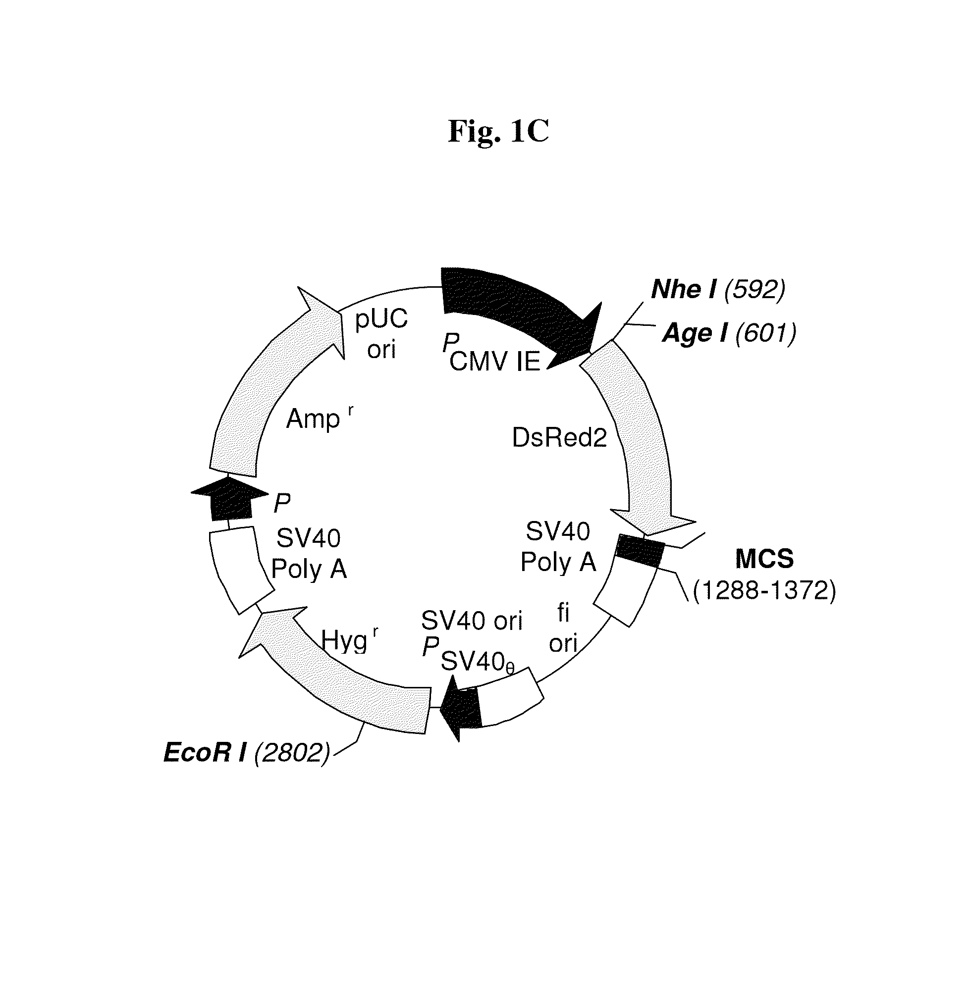
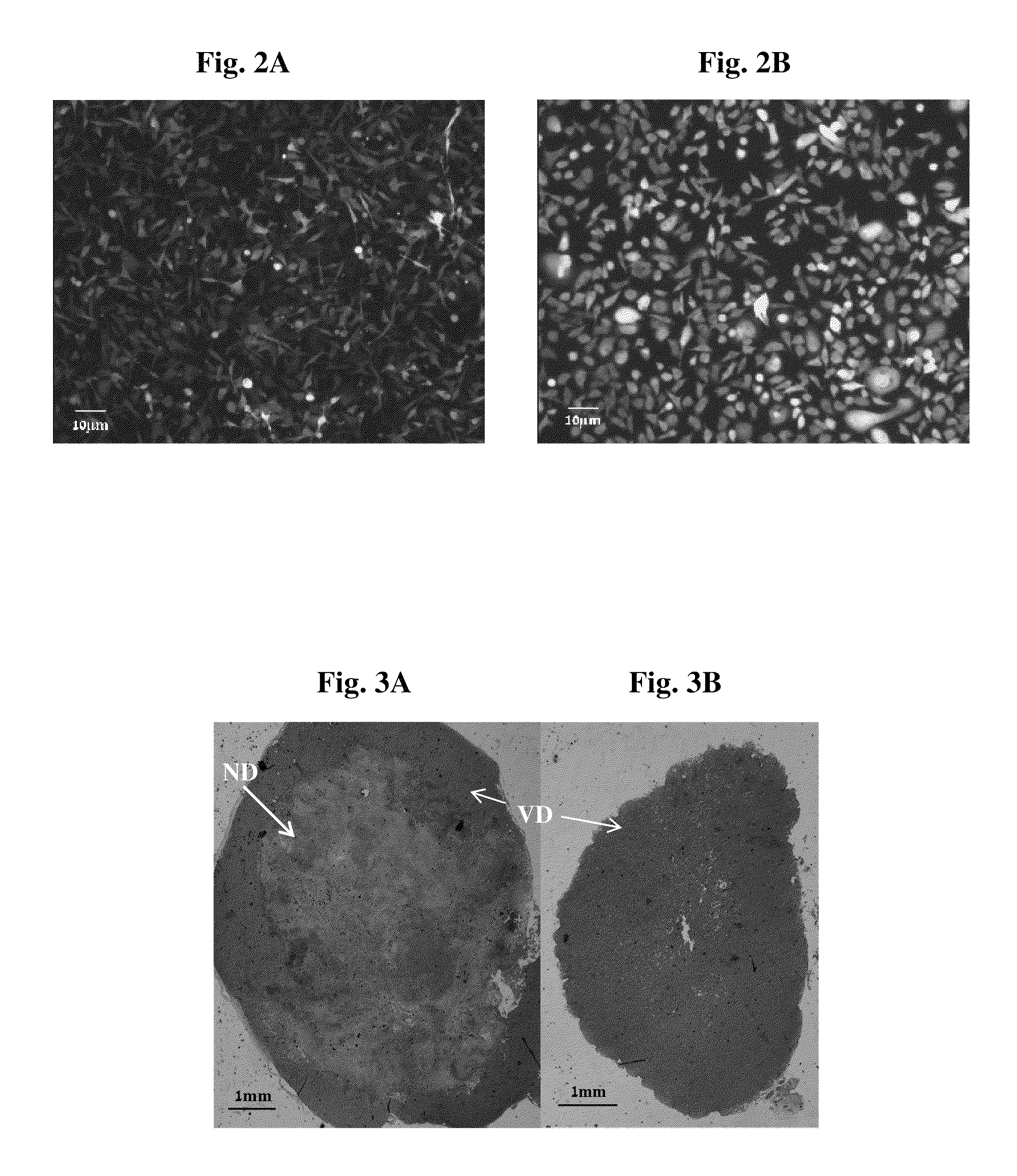
[0241]The transfection procedure was conducted in order to create a cell line that expresses RFP in a stable manner. Such a cell line can be detected by fluorescent microscopy and other fluorescence imaging means in vivo and in vitro (tissues / cells). The human breast cancer MDA-MB-231 cell line, known to generate spontaneous central necrosis, was chosen for this purpose. Two plasmids (FIGS. 1A, 1C) were used and stable clones were obtained and detected by fluorescence microscope (see FIGS. 2A-2B). The clones generated from the modified pDsRed-Monomer-Hyg-C1 plasmid (FIG. 1C) presented a stronger fluorescence. Clone 3 (FIG. 2B), transfected with modified pDsRed-Monomer-Hyg-C1, was chosen for further use.
[0242]The transfected cells expressed RFP constitutively with no reduction in the fluorescence intensity over time both in vitro and in vivo. Untransfected cells had no red auto fluorescence.
example 2
Necrotic Tumor Model—Histopathological Analysis
[0243]In order to verify that MDA-MB-231-RFP cells generate a suitable necrotic model, MDA-MB-231-RFP tumors were allowed to develop into two sizes. Histological and histopathological analysis was carried out as described in Materials and Methods section (xi). Results are demonstrated in FIGS. 3A and 3B. Large tumors of ˜1 cm3 showed a very notable necrotic domain (FIG. 3A) whereas small tumors of ˜0.5 cm3 showed no necrotic domain (FIG. 3B).
example 3
In-Vivo Fluorescence Imaging of Compound 13 Up-Take in Primary Necrotic MDA-MB-231-RFP Xenograft Tumors
[0244]The accumulation pattern of compound 13 in necrotic MDA-MB-231-RFP tumors (≧1 cm3) in vivo was examined. FIGS. 4A-4B and 5A-5B illustrate the accumulation of the fluorescence signal of compound 13 in orthotopic human breast MDA-MB-231-RFP primary tumor in the mammary pad of CD-1 nude female mice, using the Xenogen IVIS® System. Whole animal images were recorded concomitantly, using the filter sets as described in Materials and Methods section (vii) above. Dynamic fluorescence images were acquired every 1-1.5 h for 9 h, and at 24 h post injection of compound 13 (FIGS. 4A-4B), and then for every 24 h for the next 7 days (FIGS. 5A-5B). Shortly after injection of the compound 13, NIR fluorescence from the entire animal body could be detected, reflecting a high drug concentration in the circulation. Rapid clearance from the circulation, accompanied by accumulation in the liver, an...
PUM
| Property | Measurement | Unit |
|---|---|---|
| time | aaaaa | aaaaa |
| diameter | aaaaa | aaaaa |
| diameter | aaaaa | aaaaa |
Abstract
Description
Claims
Application Information
 - R&D
- Intellectual Property
- Life Sciences
- Materials
- Tech Scout
- Unparalleled Data Quality
- Higher Quality Content
- 60% Fewer Hallucinations
Browse by: Latest US Patents, China's latest patents, Technical Efficacy Thesaurus, Application Domain, Technology Topic, Popular Technical Reports.
© 2025 PatSnap. All rights reserved.Legal|Privacy policy|Modern Slavery Act Transparency Statement|Sitemap|About US| Contact US: help@patsnap.com


