RGD-(bacterio)chlorophyll conjugates for photodynamic therapy and Imaging of Necrotic tumors

a technology of rgd chlorophyll and conjugates, applied in the field of oncology, can solve the problems of poor prognosis of cancer patients, tumor aggressiveness, hypoxic and eventually necrotic, and out-growth of oxygen supply in cancer patients

Active Publication Date: 2011-03-17
YEDA RES & DEV CO LTD
View PDF4 Cites 12 Cited by
  • Summary
  • Abstract
  • Description
  • Claims
  • Application Information

AI Technical Summary

Problems solved by technology

Necrosis and hypoxia of primary and metastatic tumors have been strongly correlated with tumor aggressiveness and poor prognosis in cancer patients.
Solid tumors that reach a certain size, out grow their oxygen supply and become hypoxic and eventually necrotic.
Central necrosis, which is a common feature of invasive breast cancer, was associated with poor outcome and tumor aggression.
2003), making the identification of an area as necrotic by gene expression quite complicated.
Necrotic and hypoxic conditions are known to create a major problem in cancer therapy.
Hypoxic tumor domains are relatively resistant to radiation treatment since there is a poor promotion of the radiation assault and since stem cells that may eventually be present in the tumor volume do not respond well to the treatment, resulting in tumor re-growth (Brown et al., 1998; Dean et al., 2005).
Since most chemotherapeutic reagents impose cell death due to interactions with cycling cells, cell arrest because of hypoxia results in resistance to conventional chemotherapy, leaving non-proliferating or slow proliferating cells unharmed (Tannock, 1978).
Furthermore, hypoxic conditions usually create an acidic environment that might change the nature of the drug, making it less active (Tannock et al., 1989).
One of the more problematic aspects of solid tumors chemotherapy involves the trafficking of therapeutic agents into the tumors and especially to hypoxic and necrotic domains.
However, because of the non-regular vascularisation, many of the tumor cells are at higher distances from capillaries than cells in the normal tissues, reflected in having insufficient concentrations of antitumor agents at the cell sites.
Moreover, the enhanced interstitial fluid pressure due to the lack of lymphatic drainage reduces the convection uptake and further inhibits the distribution of drugs into the tumor cells, particularly that of macromolecules (Minchinton et al., 2006).
Histopathology and immunohistochemistry are commonly used for identification of necrosis and hypoxia; however, they are invasive and do not enable detecting in situ.
The switch to an angiogenic phenotype may represent an imbalanced expression of angiogenic factors and angiogenesis inhibitors.
The major drawback reported in the literature is the insufficient concentration of the reporting element at the site of tumors under 4-5 mm.
However, if one of these components is absent no detection is possible.
The main disadvantages of the luciferase / luciferin method are the low anatomic and image resolutions thus requiring a substantial amount of time to collect sufficient photons to form an image from an anesthetized animal.
Additionally, ex-vivo experiments are difficult to perform since ATP is required for the enzyme activity.
Importantly, the method involves subjective parameterization that reduces its quantitative value.
The main disadvantages of this method are that (1) the excitation and emission lights are prone to attenuation with increased tissue depth and (2) the autofluorescence of non labeled cells increases noise (Lyons, 2005).
There is a significant chance of transforming non-invasive DCIS lesions into an invasive, life-threatening disease if it is not treated at an early stage.
However, recurrence rates following BCS, both ipsilaterally (same breast) or contralaterally (other breast), even when accompanied by RT, were recently found to be significantly higher than after mastectomy, particularly for patients at the age of ≦40 (regression rate of 25-35%; Bijker N et al., 2006; Cutuli et al., 2002) Furthermore, multifocal lesions pose a difficulty for partial dissection and the same is true for persistently involved margins that were found critical to complete tumor regression (Cellini et al., 2005).
Additionally, the physical and psychological burden and the possible cosmetic outcomes of lumpectomy followed by RT are significant.
These drawbacks make the treatment and management of DCIS today controversial issues in breast cancer therapy and have stimulated the search for new and / or complementary modalities of treatment and prognosis.
Close to excision (less than 1 mm) or positive margins, high-grade and / or comedo necrotic areas are associated with greater risk for recurrence.
Yet, a growing body of evidence indicates deficiencies in the antiangiogenic approach.
However, the current use of MRI is limited by the available short integration time of contrast agents that shortly reside but do not selectively taken up by the tumor tissue.
Detecting tumor margins and necrosis present up to-date, two of the most challenging issues in tumor treatment.

Method used


Image

  • RGD-(bacterio)chlorophyll conjugates for photodynamic therapy and Imaging of Necrotic tumors
  • RGD-(bacterio)chlorophyll conjugates for photodynamic therapy and Imaging of Necrotic tumors
  • RGD-(bacterio)chlorophyll conjugates for photodynamic therapy and Imaging of Necrotic tumors

Examples


example 1

Transfection of Tumor Cells with Fluorescence Proteins

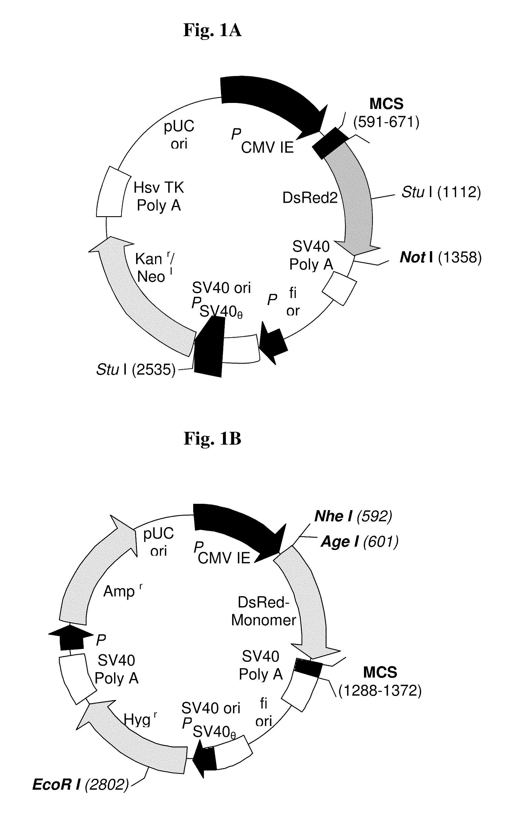
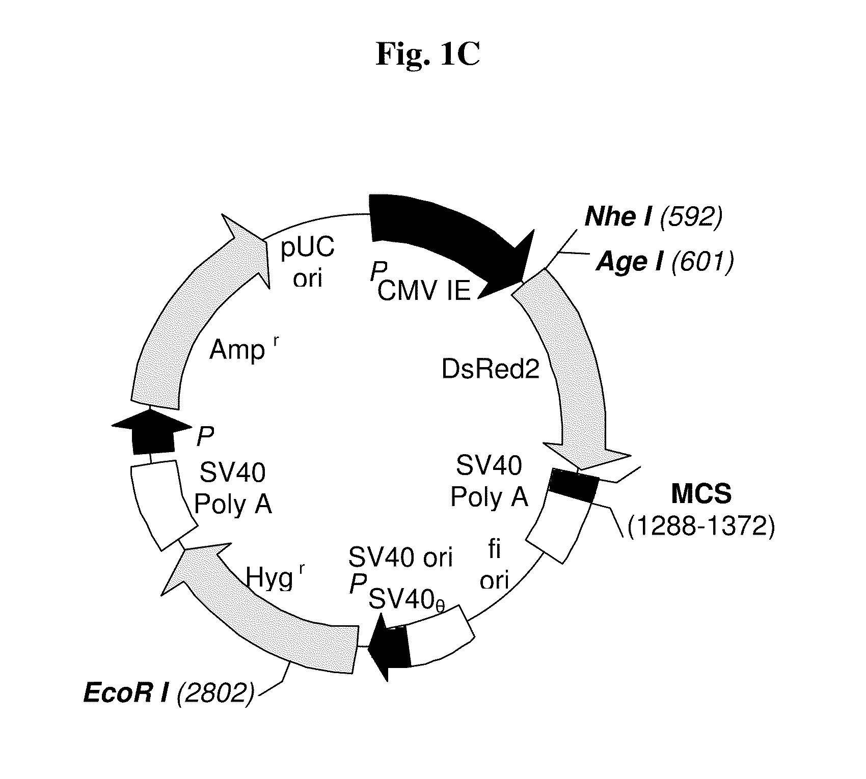
[0231]The transfection procedure was conducted in order to create a cell line that expresses RFP in a stable manner. Such a cell line can be detected by fluorescent microscopy and other fluorescence imaging means in vivo and in vitro (tissues / cells). The human breast cancer MDA-MB-231 cell line, known to generate spontaneous central necrosis, was chosen for this purpose. Two plasmids (FIGS. 1A, 1C) were used and stable clones were obtained and detected by fluorescence microscope (see FIGS. 2A-2B). The clones generated from the modified pDsRed-Monomer-Hyg-C1 plasmid (FIG. 1C) presented a stronger fluorescence. Clone 3 (FIG. 2B), transfected with modified pDsRed-Monomer-Hyg-C1, was chosen for further use.

[0232]The transfected cells expressed RFP constitutively with no reduction in the fluorescence intensity over time both in vitro and in vivo. Untransfected cells had no red auto fluorescence.

example 2

Necrotic Tumor Model—Histopathological Analysis

[0233]In order to verify that MDA-MB-231-RFP cells generate a suitable necrotic model, MDA-MB-231-RFP tumors were allowed to develop into two sizes. Histological and histopathological analysis was carried out as described in Materials and Methods section (xi). Results are demonstrated in FIGS. 3A and 3B. Large tumors of ˜1 cm3 showed a very notable necrotic domain (FIG. 3A) whereas small tumors of ˜0.5 cm3 showed no necrotic domain (FIG. 3B).

example 3

In-Vivo Fluorescence Imaging of Compound 13 Up-Take in Primary Necrotic MDA-MB-231-RFP Xenograft Tumors

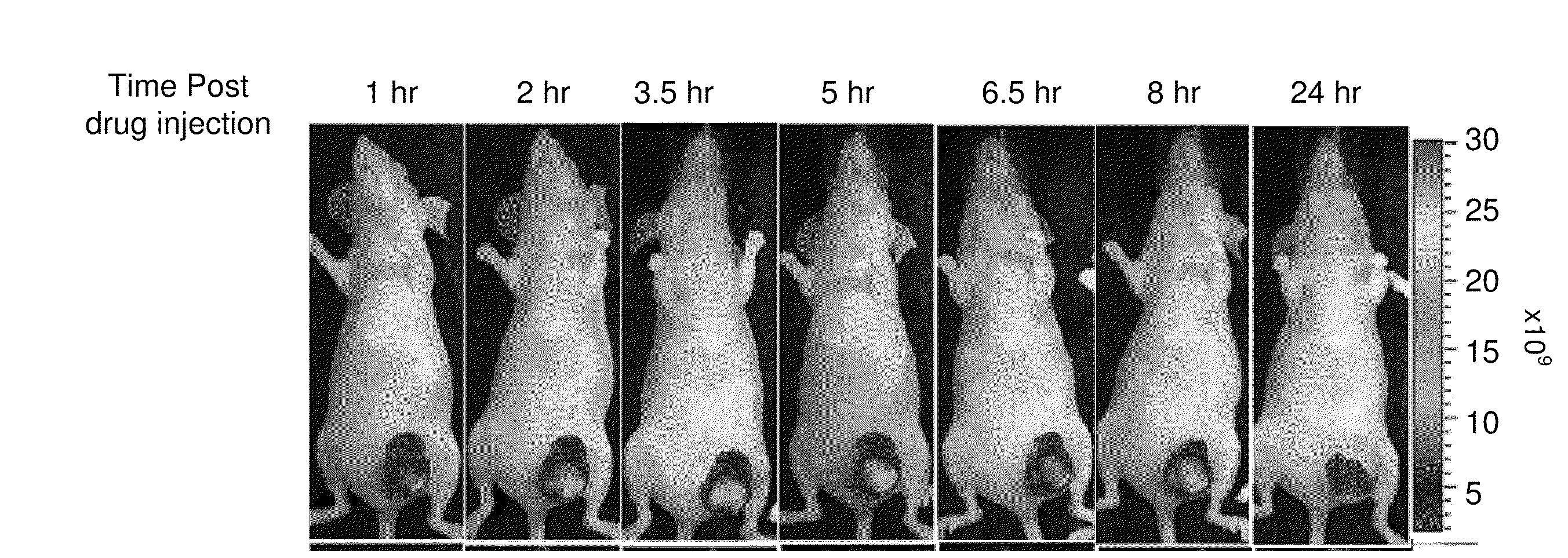
[0234]The accumulation pattern of compound 13 in necrotic MDA-MB-231-RFP tumors (≧1 cm3) in vivo was examined. FIGS. 4A-4B and 5A-5B illustrate the accumulation of the fluorescence signal of compound 13 in orthotopic human breast MDA-MB-231-RFP primary tumor in the mammary pad of CD-1 nude female mice, using the Xenogen IVIS® System. Whole animal images were recorded concomitantly, using the filter sets as described in Materials and Methods section (vii) above. Dynamic fluorescence images were acquired every 1-1.5 h for 9 h, and at 24 h post injection of compound 13 (FIGS. 4A-4B), and then for every 24 h for the next 7 days (FIGS. 5A-5B). Shortly after injection of the compound 13, NIR fluorescence from the entire animal body could be detected, reflecting a high drug concentration in the circulation. Rapid clearance from the circulation, accompanied by accumulation in the liver, an...


PUM

PropertyMeasurementUnit
Timeaaaaaaaaaa
Timeaaaaaaaaaa
Timeaaaaaaaaaa

Abstract

RGD-chlorophyll and RGD-bacteriochlorophyll conjugates that home and accumulate in necrotic tumor domains much longer than in tumor non-necrotic domains are provided for use in minimally invasive tumor-targeted imaging, tumor-targeted photodynamic therapy, and / or on-line prognosis of necrotic tumors.

Description

TECHNICAL FIELD[0001]The present invention is in the field of oncology and relates to detection of necrotic domains of tumors by tumor-targeting photodynamic imaging and treatment of said tumors by tumor-targeting photodynamic therapy using photosensitizers, particularly conjugates of chlorophyll and bacteriochlorophyll derivatives with peptides containing the RGD motif or RGD peptidomimetics.DEFINITIONS AND ABBREVIATIONS[0002]Bchl a: bacteriochlorophyll a: pentacyclic 7,8,17,18-tetrahydroporphyrin with a 5th isocyclic ring, a central Mg atom, a phytyl or geranylgeranyl group at position 173, a COOCH3 group at position 132, an H atom at position 132, methyl groups at positions 2, 7, 12, 18, an acetyl group at position 3, and an ethyl group at position 8, herein compound 1; Bphe: bacteriopheophytin a (Bchl in which the central Mg is replaced by two H atoms); Bpheid: bacteriopheophorbide a (the C-172-free carboxylic acid derived from Bphe without the central metal atom); Chl: chloroph...

Claims


Application Information

Patent Timeline
IPC IPC(8): A61K51/08A61K49/00A61K49/14A61K38/00A61P35/00A61P43/00
CPCA61K49/0036A61K49/0056A61K49/085A61K49/14A61K51/0485A61K51/088A61K51/082A61K51/044A61K51/0446A61K51/0451C07D257/04A61K41/0071A61K41/0057A61P35/00A61P35/04A61P43/00
Inventor SCHERZ, AVIGDORGOLDSHAID, LIATSALOMON, YORAM
Owner YEDA RES & DEV CO LTD
Why Eureka
Try Eureka