
Devices for separating, mixing and concentrating magnetic particles with a fluid

Active Publication Date: 2012-05-29
BIOMERIEUX BV
View PDF7 Cites 5 Cited by
  • Summary
  • Abstract
  • Description
  • Claims
  • Application Information

AI Technical Summary

Benefits of technology

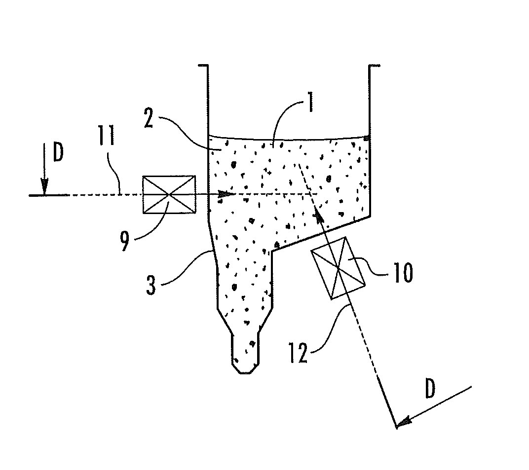
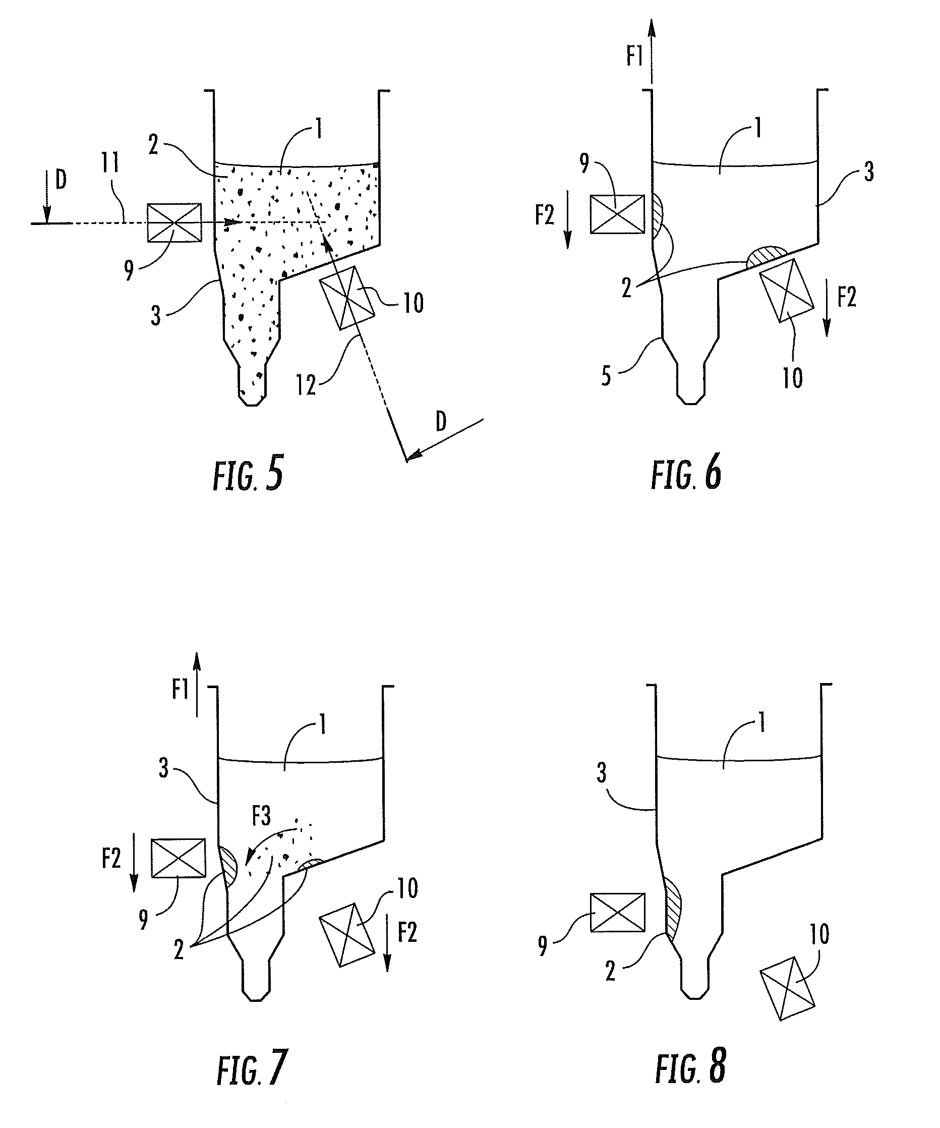
[0022]The applicant has already filed an international application WO-A-0 1 / 05510 which proposes a solution to improve the mixing. It relates to a method and device, which allows efficient mixing of magnetic or magnetizable particles in a fluid and optionally separation of the particles from said fluid. Use is made of magnetic field of opposite and changing directions. It has been found that, when magnetic or magnetizable particles in a fluid are subjected to these magnetic fields, the particles are, under the influence of the filed, efficiently contacted with the fluid. Such particles normally may tend to form a clot, which can prevent efficient mixing with a fluid. It has been found that, by subjecting the container in which the fluid and the particles are comprised, to magnetic fields of different and changing directions, the particles are efficiently separated from each other and drawn trough the fluid in such a way that a very efficient mixing process occurs. The method allows efficient mixing of particles with even very small fluid volumes. The method of the invention therefore has the advantage that it may save in, for example, washing fluids and may allow the reduction of the volume of fluid needed. Thus, for example in isolation procedures, the method of the invention allows the purification of reagents in high concentrations. Beside, whereas prior art methods can be laborious and time consuming; the method is fast and easy to perform.
[0025]In addition, in order to increase the sensitivity and the specificity of a test for demonstrating an analyte, it is sometimes necessary to reduce the volume of the buffer in which the copies of the analyte being sought are found, while at the same time conserving said analyte in its entirety.
[0032]A real need therefore exists for a method and a device for treating analytes bound to magnetic particles while at the same time conserving the amount of analyte present at the start, for example in order to increase the sensitivity and the specificity of diagnostic tests and of any chemical reaction directed towards the analyte, and to overcome the abovementioned drawbacks.
[0094]With “mixing” in this context is meant that the particles and the fluid are brought in close contact. The word “mixing” thus means contacting in a very efficient manner, such as when particles would be washed or reacted with components present in the fluid. Mixing, in this context, does not necessarily provide a homogeneous mixture after the process is finished. The particles may, when the magnets are removed, segregate to the bottom of the container in which they are comprised or may be held to the wall of the container in a particular location by the magnets. The mixing process can for example be used to wash the particles or to react the particles with a component of the liquid, or to bind a component of the liquid to a reagent coated on the particles. Likewise, the mixing process may result in the elution of a certain component originally present on the particles into the surrounding liquid. The method of the invention is applicable in each of these processes and provides an efficient, rapid and convenient way of contacting magnetic or magnetizable particles with a volume of a certain fluid.

Problems solved by technology

During an immunoassay, the fluid level in the reaction vessels after adding the required reagents is not necessarily uniform.
Known devices for separating magnetic particles have the disadvantage of requiring a relatively long time before all magnetic particles are separated from the liquid phase.
Separation time may be considerable, particularly for larger volumes.
This device has the disadvantage of requiring a relatively large number of permanent magnets to speed up separation.
If these documents can be considered for the separation of magnetic particles from a liquid of interest, they cannot authorize a performant mixing that could efficiently wash the particles and give them all chance to bind onto the magnetic particle's surfaces.
In general, this washing and elution process may be hampered by the aggregation or clogging of the magnetic particles either caused by the adsorption of specific components in the lysed sample (e.g. genomic DNA) or by residual magnetic dipole fields induced in the particles.
In particular, the use of silica coated (magnetic) particles with samples that contain significant amounts of genomic DNA (whole blood, sputum, tissue), results in a tight pellet that is difficult to process.
These methods however are difficult to automate, and / or give risk of sample to sample contamination by aerosol generation or they may degrade the nucleic acid target.
Furthermore, these methods are not well suited for very small volumes of liquid (typically 0.01 ml) as may be required for the elution process.
Such particles normally may tend to form a clot, which can prevent efficient mixing with a fluid.
Beside, whereas prior art methods can be laborious and time consuming; the method is fast and easy to perform.
These techniques require transfers of solutions and manipulations of the analyte, which lead to an inevitable decrease in the amount of analyte that can be analysed.
In addition, there are problems of evaporation and of adsorption to the walls of the containers during these manipulations.
In the case of a low concentration of analyte in the starting sample, this may cause the complete disappearance of the analyte or a decrease in the amount thereof such that it may become undetectable.
Besides the abovementioned drawbacks, these manipulations are expensive in terms of material and take a lot of time.
This remains a constant problem for many industrial applications, for example the detection of pathogenic micro organisms in a biological specimen or an industrial sample.
However the separation is not efficiently treated, especially if the magnet is of a smaller size compared to the first container, and the mixing is even not discussed.
Thus none of the above-described documents presents a process offering the advantages of separating, mixing and concentrating molecules of interest from a liquid sample in only one container.
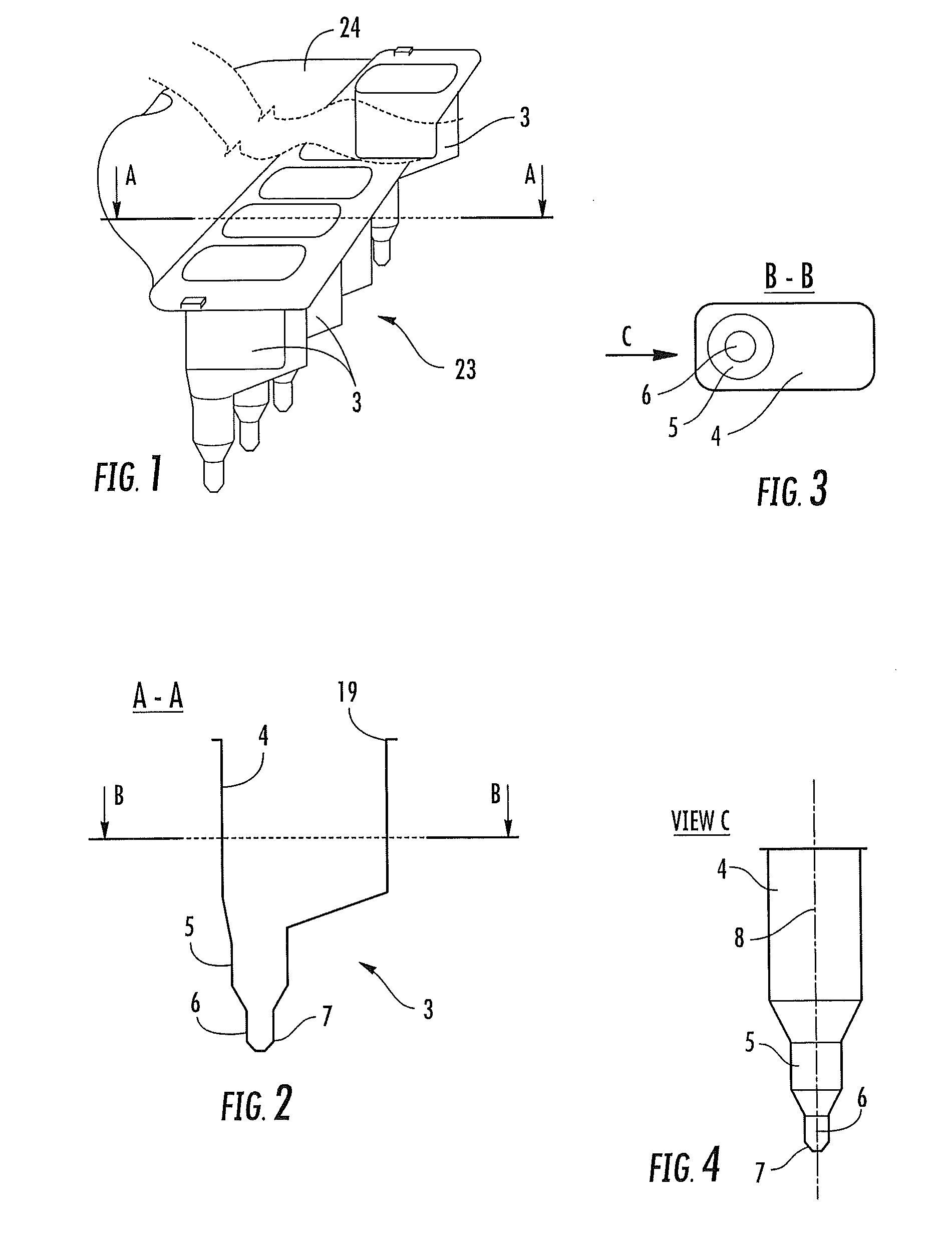
Likewise, they do not propose a container having technical features authorizing the running of such a process.
Mixing, in this context, does not necessarily provide a homogeneous mixture after the process is finished.

Method used


Image

  • Devices for separating, mixing and concentrating magnetic particles with a fluid
  • Devices for separating, mixing and concentrating magnetic particles with a fluid
  • Devices for separating, mixing and concentrating magnetic particles with a fluid

Examples


— example 1

2—EXAMPLE 1

Concentration of Ribosomal RNA from Plasma Samples

2.1—Materials and Methods

[0164]Target RNA is extracted from Escherichia coli cells using the Qiagen RNA / DNA maxi kit (article code 14162, Qiagen, Hilden, Germany). For each sample, 2 micrograms (μg) are used as input.

[0165]Sample is constituted by normal EDTA / citrate plasma samples from a pool of 100 individual blood donations.

[0166]The RNA quantification is performed using a luminescent marker for RNA (Sybr Green-II, supplied by Molecular Probes: article S-7564) in combination with a luminescent-reader for microtiter plates (“Victor2”, supplier Wallac Oy, Turku, Finland: 1-420-multilabel counter).

[0167]Purity of the output buffer is determined from the relative value for the spectral absorption at 260 nm and 280 nm (A260 / A280) using an UV spectrophotometer (supplier Unicam, Cambridge, Great Britain: Unicam UV-I).

2.2—Sample Preparation

[0168]A set of twenty-four plasma samples is prepared as follows:[0169]With each sample, ...

— example 2

3—EXAMPLE 2

Concentration of Plasmid DNA from Plasma Samples

3.1—Materials and Methods

[0181]Target is a plasmid DNA, pBR322 (article N3033L, New England Biolabs Inc., Beverly, Mass.), linearized by BamHl (article ElOlOWH, Amersham Bioscience Corp, NY, USA). Six μg of DNA is used as input per sample

[0182]The sample is constituted by 0.1 ml normal EDTA / citrate plasma from blood donations

3.2—Sample Preparation

[0183]Twenty-four samples are processed simultaneously, using the same procedure as with example 1. The individual plasma samples are added to the reaction vessels. Next, 2 ml of NucliSens lysis buffer are added. Subsequently, the plasmid DNA is spiked to the sample and 1 mg of magnetic particles is added to the mixture.

3.3—Extraction Method

[0184]The collection step, washing steps and elution step of the extraction procedure are identical to example 1.

3.4—Result

[0185]After completing the procedure, the amount of extracted DNA is determined from the optical density of the output buff...

— example 3

4—EXAMPLE 3

Recovery of Viral RNA from Sputum Samples

4.1—Materials and Methods

[0189]In this experiment, the recovery of HIV RNA that is spiked to nine sputum samples is determined using the NucliSens EasyQ HIV-I assay version 1.1 (article 285029, bioMerieux B.V., Boxtel, The Netherlands).

[0190]The RNA is HIV RNA obtained from a cultured HIV-I type B virus stock (HXB2) that is lysed using the NucliSens lysis buffer and calibrated against the WHO International HIV-I RNA standard.

[0191]The sputum samples were obtained from the University Hospital of Antwerpen (Belgium).

4.2—Sample Preparation

[0192]With each sputum sample a volume of 1 ml is mixed with 0.5 ml of protease solution (100 mg / ml) and incubated on a plate shaker for 20 minutes at room temperature to obtain a liquefied sample.

[0193]The target HIV RNA is spiked to a tube containing 2 ml of NucliSens lysis buffer, using 30.000 copies input for each sample. Together with the HIV RNA, a calibrator RNA that is part of the EasyQ kit i...


PUM

PropertyMeasurementUnit
Angleaaaaaaaaaa
Angleaaaaaaaaaa
Angleaaaaaaaaaa

Abstract

The present invention is related to devices for manipulating magnetic particles that are suspended in a fluid, possibly containing a biological entity of interest, the magnetic particles being able to bind the entity of interest, the fluid being contained in a reaction vessel constituted by a large upper compartment with a funnel shape, an elongate lower compartment with a substantially constant cross-section and a closed base. The devices are especially useful in methods for the extraction of nucleic acid to enable them for further processing.

Description

RELATED APPLICATIONS[0001]This application is a divisional of U.S. patent application Ser. No. 11 / 572,574, filed Apr. 3, 2007, now U.S. Pat. No. 7,972,516, which is a 35 USC 371 national phase application of PCT / EP2005 / 008073, filed Jul. 22, 2005, which claims the benefit of European Application Serial No. 04077153.7, filed Jul. 26, 2004, the contents of which are hereby incorporated by reference as if recited in full herein.FIELD OF THE INVENTION[0002]This invention relates to the use of magnetic or magnetizable particles, and, in particular, to methods of separating, mixing and concentrating magnetic or (super) paramagnetic particles efficiently with a fluid and optionally followed by resuspension of the particles in another fluid. The invention further provided a device for doing the same. In the below-exposed invention, the magnetic particles, paramagnetic particles and superparamagnetic particles will be called magnetic particles.BACKGROUND[0003]In many methods of biological an...

Claims


Application Information

Patent Timeline
IPC IPC(8): B03C1/02G01N1/34G01N1/38
CPCB01L3/5085B03C1/01B03C1/0332B03C1/288G01N35/0098B01L2200/025B01L2200/0647B01L2300/0858G01N1/34B03C2201/26B03C2201/18
Inventor KREUWEL, HERMANNUS JOHANNES MARIAVERWIMP, EMIEL GEREBERN MARIABEERLING, BERNARDUS JOZEF MARIASPEE, FRANCISCUS GERARDUS
Owner BIOMERIEUX BV
Learn More