Soluble epidermal growth factor receptor-like proteins and their uses in cancer detection methods
a technology receptor, which is applied in the field of soluble epidermal growth factor receptorlike proteins and their use in cancer detection methods, to achieve the effects of inhibiting ligand degradation, increasing or decreasing the half-life of egfr/erbb1, and increasing or decreasing the half-life of egfr/erbb1
- Summary
- Abstract
- Description
- Claims
- Application Information
AI Technical Summary
Benefits of technology
Problems solved by technology
Method used
Image
Examples
example i
Isolation and Characterization of Human EGFR / ERBB1 cDNAs Encoding Soluble EGFRs
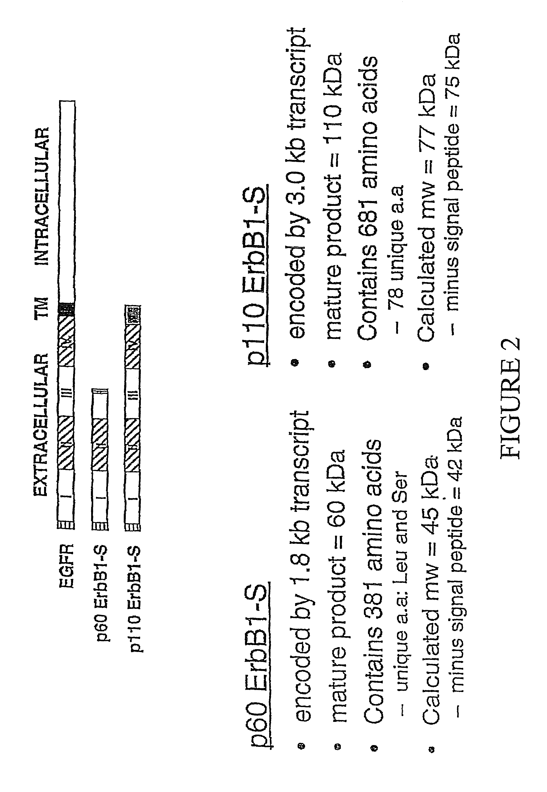
[0098]To isolate human EGFR clones that lack sequences encoding the cytoplasmic domain, but have the extracellular domain, differential hybridization was employed to screen an oligo-dT primed human placental cDNA library (Clontech, cat. # H1144x). The library was screened for clones that were positive for a ligand binding domain (LBD) specific probe (positions 174-2105), but negative for a kinase domain (KD) probe (see FIG. 2 for full length / p110 comparison).
[0099]The ligand binding domain probe was synthesized by the PCR using pXER as a template (Chen et al., Nature, 32, 820 (1987)). The forward primer was: SEQ ID NO: 7, corresponding to nucleotide positions 174-193. The reverse primer had the sequence SEQ ID NO:8, representing base pairs 2086-2105. Nucleotide numbering is according to Ullrich et al., supra, unless stated otherwise. Amplification was performed for 35 cycles (94° C. for 1 minute; 65° C. f...
example ii
Soluble Human EGFR / ERBB1R1Gene Product
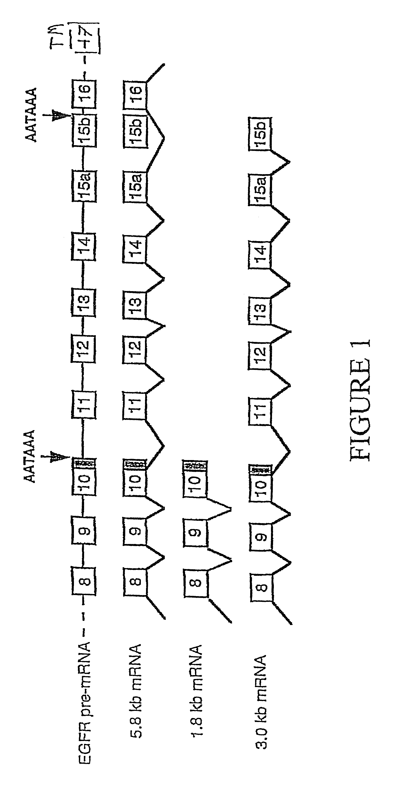
[0105]The amino acid sequence deduced from the 3.0 kb EGFR / ERBB1 cDNA, (SEQ ID NO:1), predicted a 705 amino acid polypeptide with a molecular mass of 77 kD. The first 24 amino acids code for a signal peptide; following cleavage by signal peptidases, the predicted molecular weight of this polypeptide is 75 kD. The sequence encodes subdomains I, II, III and a portion of subdomain IV of the extracellular ligand binding domain of the EGFR plus an additional 78 unique carboxy-terminal amino acids. A quail fibroblast cell line, QT6, was transiently transfected with the plasmid pDR2241, which contains the 3.0 kb EGFR / ERBB1 transcript and synthesizes a 110 kD glycosylated polypeptide (p110 sErbB1). Cells were transfected with 15 μg of pDR2241 by the calcium phosphate precipitation technique as described previously (Ausubel et al., Current Protocols in Molecular Biology, John Wiley & Sons, NY (1994)).
[0106]Transfected cells from two 10 cm plates were poo...
example iii
Growth Inhibitory Potential of Soluble ErbB1Receptors on Ovarian Carcinoma Cell Growth in Vitro
[0108]To examine the affect of p110 sErbB1 on EGFR-regulated cell growth, stable CHO cell lines expressing p110 and / or EGFR have been established and clonally isolated. Co-expression of p110 sErbB1 in cells expressing p170 EGFR results in the rapid and unexpected induction of cell rounding (24 hr) and programmed cell death (48 hr). See FIG. 5. We have verified that the mechanism of cell death is apoptotic based on nuclear morphology, and Hoechst staining. Interestingly, cell death does not occur in CHO cells treated similarly but expressing a truncated EGFR mutant that has lost most of its extracellular domain (i.e., the type III variant, originally cloned from a human glioma). These results were initially discovered using transient transfection analyses, but since that time inducible expression of p110 in CHO cells has been established using an ecdysone promoter system (Invitrogen), and h...
PUM
 Abstract
Description
Claims
Application Information
 - R&D
- Intellectual Property
- Life Sciences
- Materials
- Tech Scout
- Unparalleled Data Quality
- Higher Quality Content
- 60% Fewer Hallucinations
Browse by: Latest US Patents, China's latest patents, Technical Efficacy Thesaurus, Application Domain, Technology Topic, Popular Technical Reports.
© 2025 PatSnap. All rights reserved.Legal|Privacy policy|Modern Slavery Act Transparency Statement|Sitemap|About US| Contact US: help@patsnap.com


