C3 exoenzyme-coated stents and uses thereof for treating and preventing restenosis
a technology of c3 exoenzyme and stent, which is applied in the field of c3 exoenzymecoated stents, can solve the problems of high incidence and recurrence of constriction, and achieve the effect of facilitating cellular uptake of chimeric c3 exoenzym
- Summary
- Abstract
- Description
- Claims
- Application Information
AI Technical Summary
Benefits of technology
Problems solved by technology
Method used
Image

Examples
experiment 1
Expression and Purification of C3 Exoenzyme
[0033]C3 exoenzyme was prepared as previously described (Dillon and Feig, 1995). Competent cells of Escherichia coli strain BL21 were transformed with a vector containing a cDNA encoding a glutathione-S-transferase (GST)-C3 exoenzyme fusion protein. Transformed cells were grown to logarithmic phase, and expression of the fusion protein was induced with 200 μM isopropylthiogalactoside (IPTG) at 32° C. for 3 hours. Cell lysates were prepared and incubated with GST-Sepharose beads for 1 hour at 4° C. The beads were washed and incubated overnight at 4° C. with 3 units / ml thrombin (for cleavage of the C3 exoenzyme from the GST fusion protein), which was then removed by incubating the supernatant with antithrombin-Sepharose beads for 1 hour at 4° C. The supernatant was concentrated with a Centricon-10 (Amicon Inc, Beverly, Mass.). Protein concentration was determined by Bradford assay and the supernatant was aliquoted and frozen in liquid nitroge...
experiment 2
Testing Effects of C3-Exoenzyme-Coated Stents In Vivo
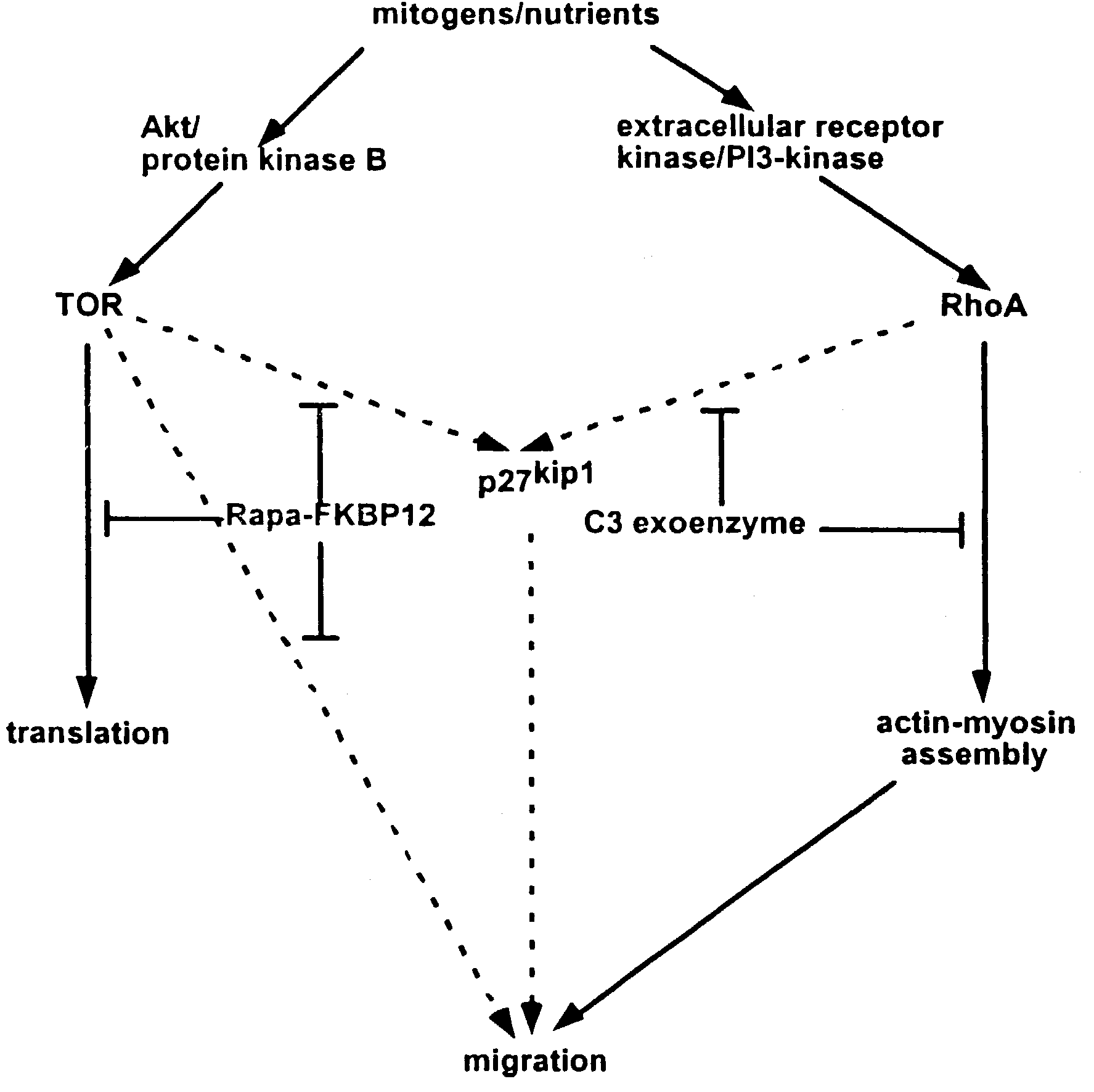
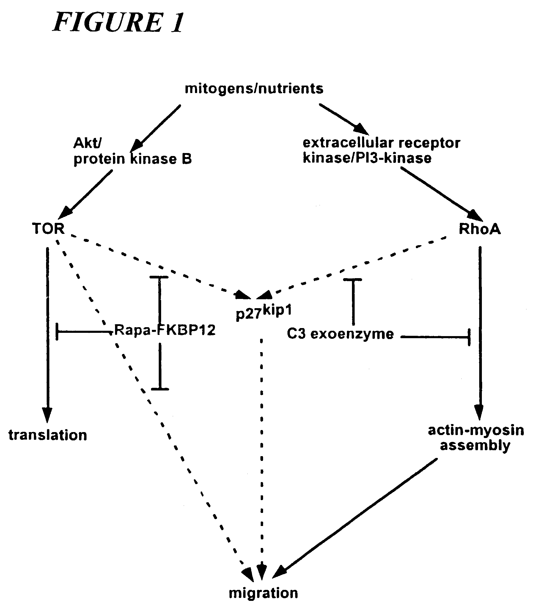
[0035]C-3 exoenzyme, like rapamycin, inhibits vascular SMC migration in vitro. To confirm the effects demonstrated in non-cellular systems, the biological effects of stents coated with the active compound are investigated in vivo by implanting stents into animal models and monitoring the occurrence of restenosis after a period of time in the animal. Such tests confirm the utility of the methods of the invention for use in mammals, are useful for obtaining data on proper dosing of the drugs, and fulfill the mandate of the U.S. Food and Drug Administration for animal testing prior to use in human subjects. Specific guidelines outlining the required tests have recently been published (Schwartz et al., 2002).
[0036]Testing of the C3 exoenzyme-coated stents is carried out in pigs which are commonly used for stent evaluation because of their anatomical (number and size of coronary arteries) and physiological similarity (blood clotting sy...
experiment 3
Surgical Procedure Used to Test Efficacy of Coated Stents In Vivo
[0043]1. One day prior to the procedure, the animals are administered aspirin (325 mg / d PO) and ticlopidine (250 mg / d), to be continued daily until the animals are euthanized.[0044]2. Ketamin (35 mg / kg) and glycopyrrolate (0.01 mg / kg) are administered intra-muscularly as premedication.[0045]3. Intubation and placement of a venous line are performed.[0046]4. The animals are anesthetized with inhaled isoflurane / oxygen.[0047]5. The right femoral groin area is shaved and sterilized.[0048]6. Cut-down to the right femoral artery or left carotid artery is carried out, followed by arteriotomy and insertion of an 8F introducer.[0049]7. An indwelling subclavian catheter is inserted to withdraw blood samples.[0050]8. For heparinization, a 10,000 U IV bolus is used, and the dosage continued at 5000 U per hour.[0051]9. Bretylium (5 mg / kg IV bolus) is administered and then infusion continued at 1 mg / min.[0052]10. An 8F hockystick co...
PUM
 Abstract
Description
Claims
Application Information
 - Generate Ideas
- Intellectual Property
- Life Sciences
- Materials
- Tech Scout
- Unparalleled Data Quality
- Higher Quality Content
- 60% Fewer Hallucinations
Browse by: Latest US Patents, China's latest patents, Technical Efficacy Thesaurus, Application Domain, Technology Topic, Popular Technical Reports.
© 2025 PatSnap. All rights reserved.Legal|Privacy policy|Modern Slavery Act Transparency Statement|Sitemap|About US| Contact US: help@patsnap.com