
TNF-α binding molecules

a technology of binding molecules and nucleic acids, applied in the field of tnf- binding molecules and nucleic acid sequences, can solve the problems of short serum half-life, inability to trigger certain human beings, limited use in vivo, etc., and achieve low dissociation rate, high binding affinity, and high association rate

Active Publication Date: 2006-09-05
TRIMER BIOTECH LLC
View PDF85 Cites 10 Cited by
  • Summary
  • Abstract
  • Description
  • Claims
  • Application Information

AI Technical Summary

Benefits of technology

[0008]The present invention provides TNF-α binding molecules and nucleic acid sequences encoding TNF-α binding molecules. In particular, the present invention provides TNF-α binding molecules with a high binding affinity, a high association rate, a low dissociation rate with regard to human TNF-α, and enhanced neutralization properties in vitro and in vivo. Preferably, the TNF-α binding molecules of the present invention comprise light and / or heavy chain variable regions with fully human frameworks. In particularly preferred embodiments, the TNF-α binding molecules of the present invention comprise light and / or heavy chain variable regions with germline frameworks (e.g. human germline frameworks).

Problems solved by technology

While these mouse anti-hTNF-α antibodies often displayed high affinity for hTNF-α and were able to neutralize hTNF-α activity, their use in vivo has been limited by problems associated with the administration of mouse antibodies to humans, such as a short serum half-life, an inability to trigger certain human effector functions, and elicitation of an unwanted immune response against the mouse antibody in a human (the “human anti-mouse antibody” (HAMA) reaction).
However, because these antibodies still retain a substantial number of murine residues, they still may elicit an unwanted immune reaction, the human anti-chimeric antibody (HACA) reaction, especially when administered for prolonged periods (see e.g., Elliott, et al., (1994) Lancet 344:1125–1127; and Elliot, et al., (1994) Lancet 344:1105–1110).
However, these hybridoma-derived monoclonal autoantibodies were reported to have an affinity for hTNF-α that was too low to measure by conventional methods, were unable to bind soluble hTNF-α and were unable to neutralize hTNF-α-induced cytotoxicity (see Boyle, et al., (1993) Cell. Immunol. 152:556–581).
However, because of their relatively fast dissociation kinetics, and relatively low affinity, these antibodies may not be suitable for therapeutic use.

Method used


Image

  • TNF-α binding molecules
  • TNF-α binding molecules
  • TNF-α binding molecules

Examples


example 1

Construction and Screening of Anti-TNF-α Fab Fragments with Synthetic CDRs and Human Frameworks

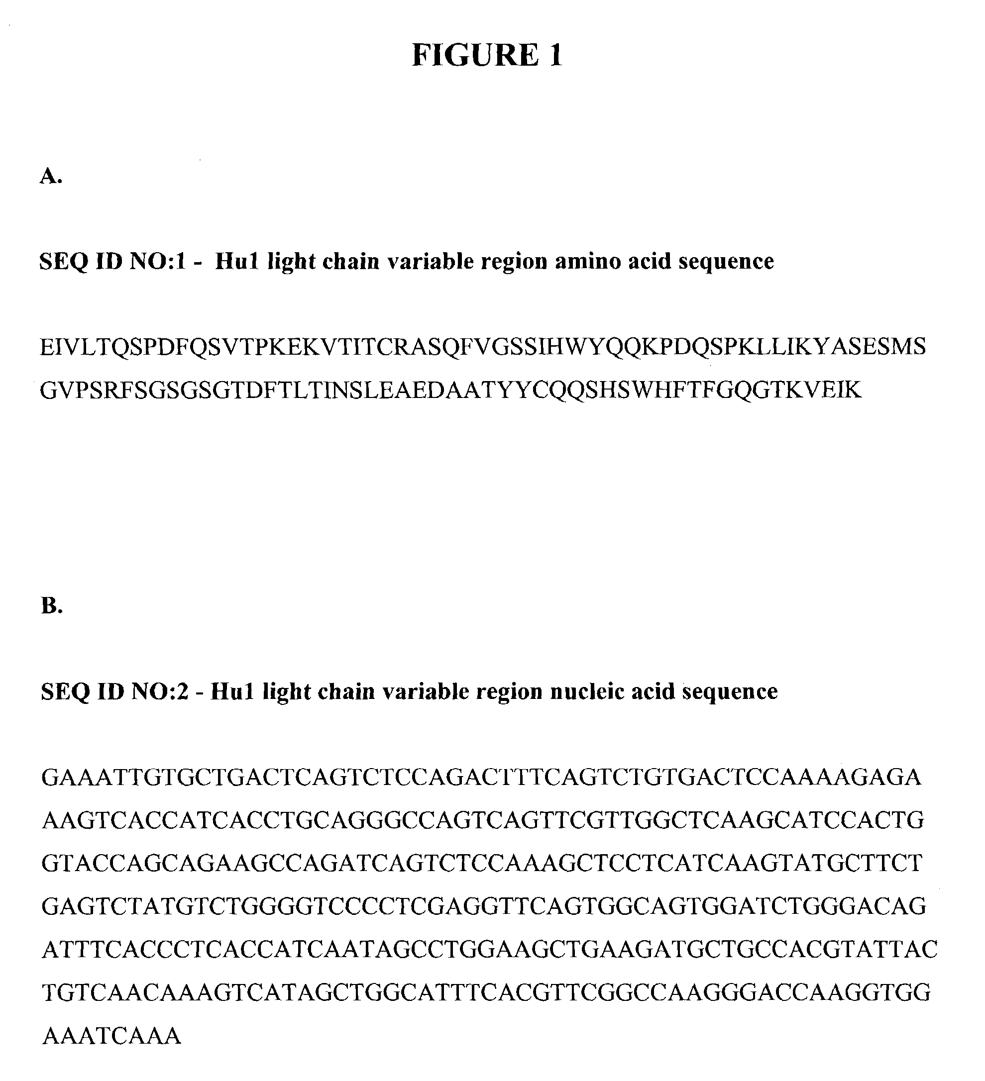
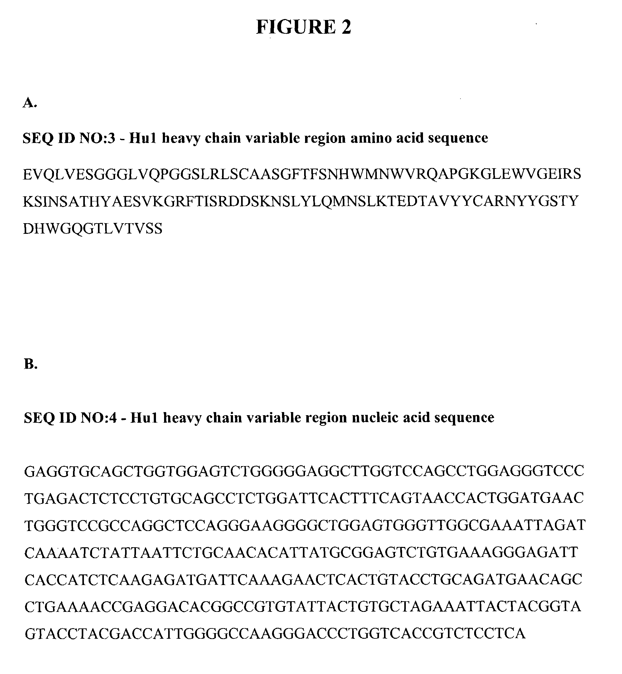
[0206]This example describes the construction of anti-TNF-α Fab fragments with fully human framework regions and synthetic CDRs. The nucleic acid sequences encoding the hu1 CDRs and framework regions are shown in SEQ ID NOs: 2 and 4, respectively. The six hu1 CDRs employed were as follows: CDRL1 (SEQ ID NO: 10); CDRL2 (SEQ ID NO: 20); CDRL3 (SEQ ID NO: 34); CDRH1 (SEQ ID NO: 36); CDRH2 (SEQ ID NO: 44); and CDRH3 (SEQ ID NO: 54).

[0207]In order to identify mutations that improve the binding properties of hu1, libraries of synthetic CDRs were inserted into a deletion template as described below. Standard mutagenesis techniques (Kunkel, T. A., (1985) Proc. Natl. Acad. Sci. U.S.A, 82: 488–492, herein incorporated by reference) were employed to individually replace each hu1 CDR with a pool of mutagenic oligonucleotides. In this example, the CDRs include residues that encompass both the Kabat and...

example 2

Characterization of Anti-TNF-α Binding Molecules with Improved Binding Properties

[0218]This example describes various in vitro testing procedures used to identify particular characteristics of the eight anti-TNF-α binding molecules from Example 1. First, the ability of these Fab fragments to prevent TNF-α-mediated cell death was assessed by evaluating their protective effects on L929 mouse fibroblasts. Specifically, L929 mouse fibroblast cells were distributed in individual wells in a 96-well plate at a density of 20,000 cells per well and maintained according to ATCC recommendations. After overnight incubation, Fab fragments released from the periplasmic space were serially diluted in culture medium, sterile filtered and 50 μl were added to the wells along with actinomycin D to a final concentration of 1 μg / ml. Immediately thereafter, hTNF-α was added to a final concentration of 80 pg / ml and the plates were placed in a humidified incubator at 37° C. in a 5% CO2 atmosphere. After ov...

example 3

In Vivo Examples with A10K

[0223]This example describes two in vivo assays with the A10K intact IgG. In particular, this example describes the in vivo TNF-α neutralizing effects of A10K in mice, as well as the protective effects of A10K on TNF-α mediated polyarthritis in mice.

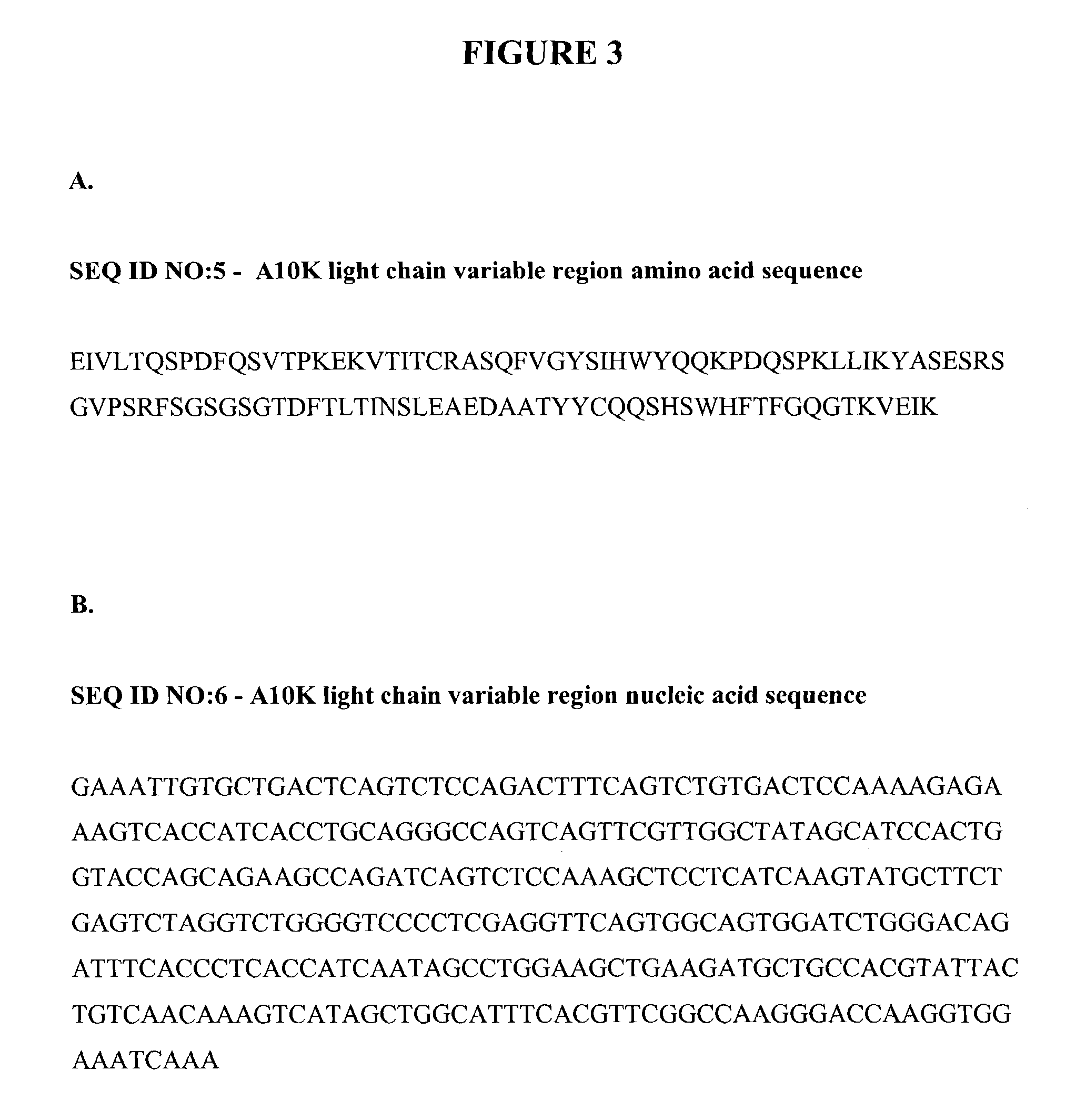
[0224]The TNF-α neutralizing ability of A10K in vivo was evaluated as follows. Female C3H / HeN mice (˜9 weeks of age) were injected intraperitoneally with recombinant human TNF-α (5 μg / mouse) in combination with the sensitizing agent galactosamine (18 mg / mouse). In untreated mice, these conditions result in approximately 90 percent mortality within 48 hours. The neutralizing effect of A10K was evaluated by injecting mice with 0.8 and 4 mg / kg, intravenously, 24 hours prior to hTNF-α challenge. A10K demonstrated a dose-dependent inhibition of mortality, with complete protection being observed at the higher dose investigated. These results are presented in FIG. 11.

[0225]The effect of A10K on TNF-α mediated polyarthr...


PUM

PropertyMeasurementUnit
Molar densityaaaaaaaaaa
Frequencyaaaaaaaaaa
Molar densityaaaaaaaaaa

Abstract

The present invention relates to TNF-α binding molecules and nucleic acid sequences encoding TNF-α binding molecules. In particular, the present invention relates to TNF-α binding molecules with a high binding affinity, a high association rate, a low dissociation rate with regard to human TNF-α and that are capable of neutralizing TNF-α at low concentrations. Preferably, the TNF-α binding molecules of the present invention comprise light and / or heavy chain variable regions with fully human frameworks (e.g. human germline frameworks).

Description

FIELD OF THE INVENTION[0001]The present invention relates to TNF-α binding molecules and nucleic acid sequences encoding TNF-α binding molecules. In particular, the present invention relates to TNF-α binding molecules with a high binding affinity, a high association rate, a low dissociation rate with regard to human TNF-α, and that are capable of preventing TNF-α mediated cytotoxicity at low concentrations. Preferably, the TNF-α binding molecules of the present invention comprise light and / or heavy chain variable regions with fully human frameworks (e.g. human germline frameworks).BACKGROUND OF THE INVENTION[0002]Tumor necrosis factor alpha (TNF-α) is a cytokine produced by numerous cell types, including monocytes and macrophages, that was originally identified based on its ability to induce the necrosis of certain mouse tumors. Subsequently, a factor termed cachectin, associated with cachexia, was shown to be identical to TNF-α. TNF-α has been implicated in the pathophysiology of a...

Claims


Application Information

Patent Timeline
IPC IPC(8): C07K16/00C07K16/24
CPCC07K16/241A61K2039/505G01N2333/525C07K2317/24
Inventor WATKINS, JEFFRY D.VASSEROT, ALAIN P.MARQUIS, DAVIDHUSE, WILLIAM D.
Owner TRIMER BIOTECH LLC
Learn More