
Morpholino imaging and therapy

Inactive Publication Date: 2005-05-31
UNIV OF MASSACHUSETTS
View PDF25 Cites 107 Cited by
  • Summary
  • Abstract
  • Description
  • Claims
  • Application Information

AI Technical Summary

Benefits of technology

[0017]Accordingly, an object of the present invention is to provide a kit and a method useful for targeting of a diagnostic or therapeutic agent in a mammal which can be prepared from relatively inexpensive starting materials but yet provides better specificity, stability, predictable targeting and / or more desirable antigen-antibody effects than conventional and other known kits and methods.
[0018]It is another object to provide an alternative method useful for tumor localization / imaging by stepwise targeting using a pair of single-stranded Morpholino oligomers (MORFs, as defined below) instead of strep / avidin-biotin, peptide nucleic acids and other oligomers, wherein a radiolabeled targeting moiety is highly accreted to the primary target-specific binding site within the target thereby providing a good tumor to non-tumor ratio for imaging purposes.

Problems solved by technology

One of the problems encountered in direct targeting methods is that a relatively small fraction of the conjugate actually binds to the target site, while the majority of the conjugate remains in circulation and compromises in one way or another the function of the targeted conjugate.
Other problems include high background and low resolution when a diagnostic agent is administered and marrow toxicity or systemic side effects when a therapeutic agent is attached to a long circulating targeting moiety.
However, difficulties have arisen in the applications of biotin-avidin or (strept)avidin system during pretargeting.
First of all, unless properly constructed, radiolabeled biotins may be subject to plasma biotinidase degradation.
A three-step strategy involving administration of biotinylated monoclonal antibody, avidin, followed by radiolabeled biotin alleviates some of the drawbacks; however, this procedure is considered complex for imaging and does not address immunogenecity.
In addition to the need to prepare bispecific antibodies, this approach may suffer from lower affinities.
In part, this may be attributed to constraints placed on these parameters by the application.
Despite the several advantages over the strept / avidin-biotin and bispecific antibody-hapten systems, a few limitations exist in the use of these oligomers in pretargeting.
These limitations include poor specificity, possible insolubility in aqueous solutions, and high costs.

Method used


Image

  • Morpholino imaging and therapy
  • Morpholino imaging and therapy
  • Morpholino imaging and therapy

Examples


example 1

Coupling of MN-14 with MORF15 AND MORF18

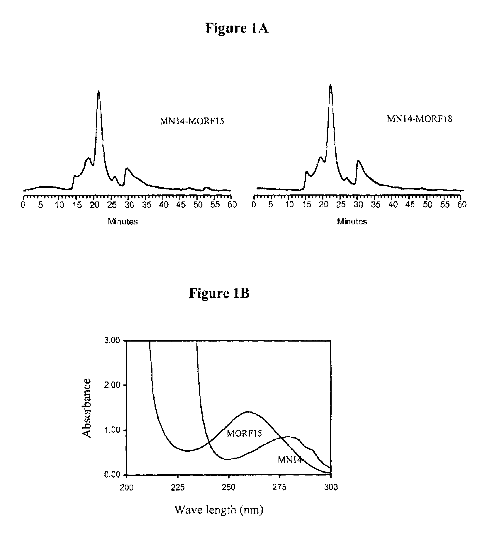
[0076]A solution of murine anti-CEA IgG antibody MN-14 (IgG1 subtype, MW 150-kDa) was prepared at a concentration of 1000 μg / 83 μL in phosphate buffered saline (pH 7.0-7.2) and was conjugated with either MORF15 (500 μg / 250 μL) or MORF18 (600 μg / 250 μL) in 2-(N-morpholino)ethane sulfonic acid (MES), pH 5.0, in the presence of 1-ethyl-3(3-dimethylaminopropyl)carbodiimide (EDC) in water (1000 μg / 50 μL). The above mixture was incubated at room temperature for at least 1 hour.

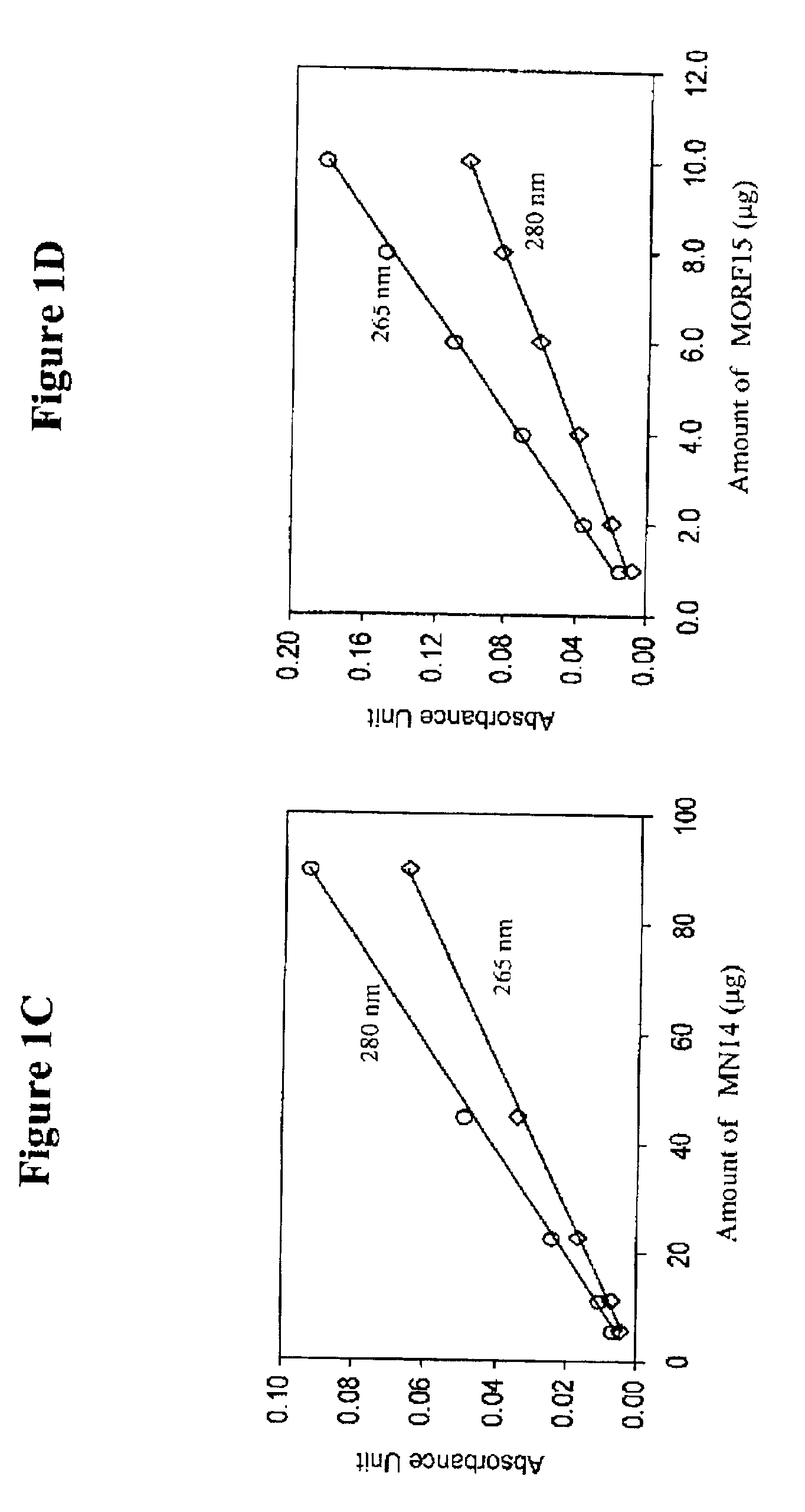
[0077]Purification was achieved on a 0.7×20 cm Sephadex G-100 column with 0.05 mol / L, pH 7.0 phosphate buffer as eluant. The concentration of the recovered fraction(s) was estimated with respect to MN14 by UV absorbance at 280 nm using the absorbance coefficient of 1.40 μL / μg.

[0078]The antibody-MORF conjugates were characterized in terms of antibody concentration and MORF group per molecule. Previously, groups per molecule were estimated by adding increasing amounts of radiola...

example 2

Coupling of cMORF15 and cMORF18 to NHS-MAG3

[0081]The conjugation of cMORF15 and cMORF18 with NHS-MAG3 was accomplished as described in Mardirossian, G. et al., J. Nucl. Med. 38:907-13 (1997) and Wang, Y. et al. Bioconj. Chem. 12:807-816 (2001). NHS-MAG3 powder (410 μg) was conjugated to both cMORF15 and cMORF18 (880 μg / 200 μL) in 0.2M HEPES, pH 8. Fifty microliters of HEPES was added to the resulting mixture and incubated for 4 hours. CMORFs (880 μg) were dissolved in 250 μl of 0.2 M HEPES buffer (pH 8.0) and were added to a vial containing 1150 μg of solid NHS-MAG3. After vortexing, the mixture was incubated at room temperature for at least 1 hr. The molar ratio of MORF18 to NHS-MAG3 was about 1:20. The incubated solution was separated on a 0.7×20 cm column of P4 with an eluant of 0.25 M ammonium acetate buffer, pH 5.2. The concentration with respect to cMORF of the recovered fractions were estimated by UV absorbance at 265 nm using an absorbance coefficient of 31 μL / μg. The peak ...

example 3

Radiolabeling of cMORF15-MAG3 and cMORF18-MAG3

[0083]Twenty-five microliters of cMORF15-MAG3 and cMORF18-MAG3 (0.25 M ammonium acetate, pH 5.2) was mixed with 6 μL sodium tartrate solution (50 mg / ml of fresh sodium tartrate in 0.5 M sodium bicarbonate, 0.25M ammonium acetate, 0.18 ammonium hydroxide, pH 9.2). This was followed by adding 20 μL of about 5 mCi of 99mTc-pertechnetate generator eluant. Finally, 2 μL tin(II) chloride (1 μg / μL in 10 mM HCl; Sigma) was quickly added by agitation. The conjugates were capped and placed in a boiling water bath for 20-30 min.

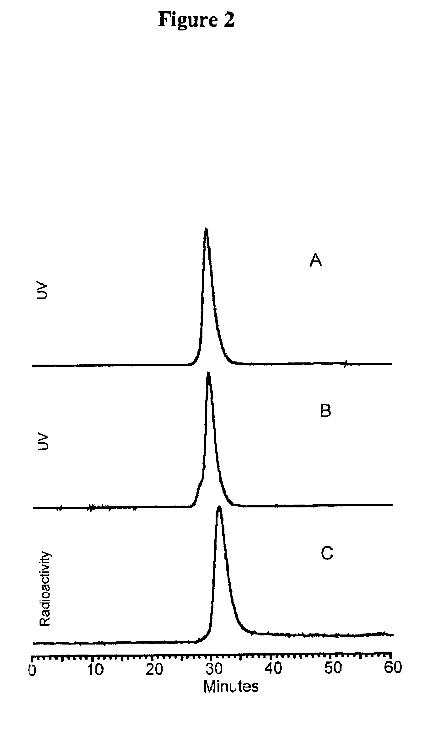
[0084]After 5-15 minutes at room temperature, the labeled-cMORFs-MAG3 was purified on a P4 column (0.7×20 cm) with 0.05 M phosphate buffer, pH 7 as the eluant. The peak was identified by counting fractions in a dose calibrator. The purified product was analyzed by size-exclusion HPLC using a Superose-12 (Pharmacia, Piscataway, N.J.). FIG. 1 represents a size exclusion HPLC radiochromatogram showing that the peak-times for ...


PUM

PropertyMeasurementUnit
Massaaaaaaaaaa
Massaaaaaaaaaa
Massaaaaaaaaaa

Abstract

The present invention provides a kit and a method for targeting of a diagnostic or therapeutic agent to a target site in a mammal having a pathological condition. The kit comprises, in separate containers, (a) a first conjugate comprising a targeting moiety and a Morpholino oligomer, wherein the targeting moiety selectively binds to a primary, target-specific binding site of the target site or to a substance produced by or associated with the target site; (b) optionally, a clearing agent; and (c) a second conjugate comprising a complementary Morpholino oligomer and a diagnostic agent or therapeutic agent. The method comprises administering (a), optionally (b), and (c) to a mammal.

Description

CROSS-REFERENCE TO RELATED APPLICATIONS[0001]This application is based on U.S. provisional patent application Ser. No. 60 / 279,809, filed Mar. 30, 2001 now abandoned, and 60 / 341,794, filed Dec. 21, 2001. The entire contents of these applications, including their specifications, claims and drawings, are incorporated herein by reference in their entirety.FIELD OF THE INVENTION[0002]The present invention is directed to a kit for targeting of a diagnostic or therapeutic agent to a target site in a mammal, as well as to a method for diagnosing or treating a pathological condition in a multistep process using a kit containing a complementary pair of single-stranded Morpholino oligomers.BACKGROUND OF THE INVENTION[0003]The objective of drug targeting research is to improve the effectiveness of therapeutic drugs by delivering them directly to the targeted tumor sites and allowing a more effective dosing at these sites, thereby reducing non-tumor-related side effects. Another objective is to ...

Claims


Application Information

Patent Timeline
IPC IPC(8): A61K39/395A61B5/055A61K51/00C12Q1/68A61KA61K31/535A61K41/00A61K45/00A61K47/48A61K49/08A61K51/04A61K51/10A61P35/04C07K16/30C12P21/08
CPCA61K47/48746C07K16/3007B82Y5/00A61K51/0495A61K47/6897A61P35/04
Inventor HNATOWICH, DONALD J.RUSCKOWSKI, MARYLIU, GUOZHENG
Owner UNIV OF MASSACHUSETTS
Learn More