Paramagnetic particles that provide improved relaxivity
a technology of relaxation rate and magnetism, applied in the field of improved contrast agents for magnetic resonance imaging, can solve the problems of disrupting statistical alignment, too slow to generate detectable amounts of energy, and left to their own devices relaxation rate of relevant hydrogen nuclei,
- Summary
- Abstract
- Description
- Claims
- Application Information
AI Technical Summary
Problems solved by technology
Method used
Image
Examples
example 1
Preparation of Contrast Agent
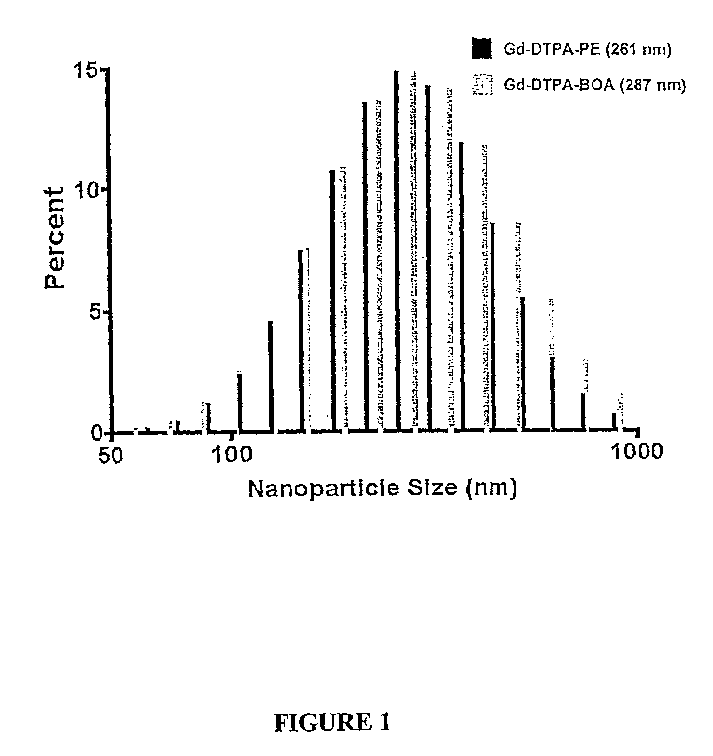
As set forth in Preparation A, Either gadolinium diethylene-triamine-pentaacetic acid-bis-oleate (Gd-DTPA-BOA; Gateway Chemical Technologies, St. Louis, Mo.) or DTPA-phosphatidylethanolamine (DTPA-PE; Gateway Chemical Technologies, St. Louis, Mo.), was included in the surfactant co-mixture at a concentration of 20 mole % of the total lipid membrane. Gadolinium chloride was added in excess proportions as a post-emulsification step to nanoparticles formulated with DTPA-PE. Unbound gadolinium was removed by dialysis on the nanoparticles against distilled deionized water (300,000 MW cut-off, Spectrum Laboratories, Rancho Dominguez, Calif.). Gadolinium-DTPA-BOA was incorporated into the surfactant lipids as the complete paramagnetic compound. Both Gd-DTPA-BOA and Gd-DTPA-PE emulsions were tested for free Gd3+ using the arsenazo III reaction and showed no sign of unbound lanthanide.
The concentration of Gd3+ was calculated from the reactants used during formula...
example 2
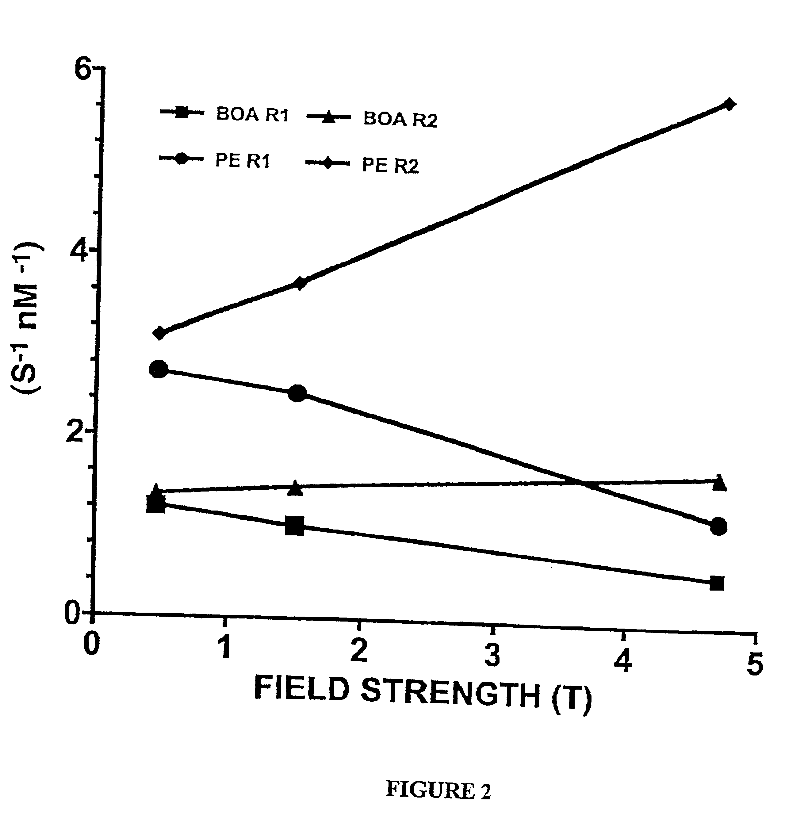
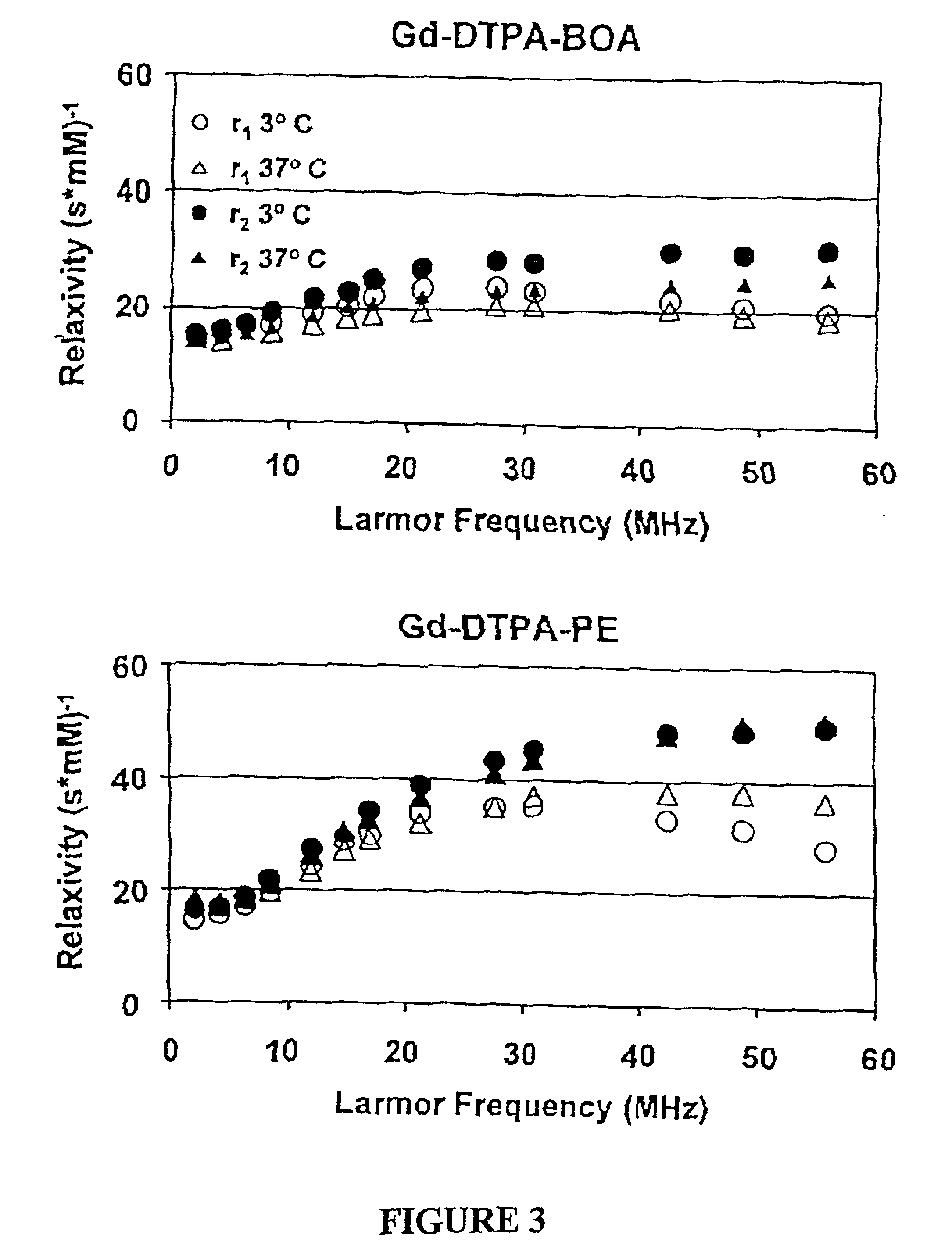
Paramagnetic Nanoparticle Sample Preparation and Assessment of T1 and T2 Relaxivities at 0.47 T, 1.5 T and 4.7 T
Gd-DTPA-BOA and Gd-DTPA-PE nanoparticles prepared in Example 1 were diluted to 0, 4, 6, 8, 10 and 12% PFOB (v / v) with distilled deionized water. The initial nanoparticle formulation contained 26.1 mol / L 19F and the diluted aliquots had 0, 3.915, 5.22, 6.525 and 7.83 mol / L 19F, respectively. Total gadolinium content was determined by neutron activation analysis. The gadolinium contents of the Gd-DTPA-BOA nanoparticle dilutions were 0; 0.336; 0.504; 0.672; 0.84; and 1.01 mmol / L Gd3+. The paramagnetic ion concentrations in Gd-DTPA-PE samples were 0; 0.579; 0.869; 1.16; 1.45; and 1.74 mmol / L Gd3+.
The proton longitudinal and transverse relaxation rates (1 / T1 and 1 / T2, respectively) of each sample were measured at 40° C. on a Bruker MQ20 Minispec NMR Analyzer with a field strength of 0.47 T. T1 was measured using an inversion recovery sequence with 10 inversion delay values, whi...
example 3
19F Spectroscopy and Imaging
The 19F signal intensities of Gd-DTPA-BOA and Gd-DTPA-PE nanoparticles were characterized at 0.47 T and 4.7 T, but the necessary RF channel was unavailable for study at 1.5 T. At 0.47 T, 19F spectra were collected from each sample and the signal was quantified with respect to a reagent-grade PFOB standard. At 4.7 T, spin echo 19F images were collected from a six chamber phantom using a 1.5 cm single turn solenoid coil, dual-tuned to 1H and 19F. The imaging parameters were: TR=5000 ms, TE=6.3 ms, number of signal averages=35, image matrix=256 by 256, FOV=2 by 2 cm, flip angle=90°, slice thickness=1 mm. The relative 19F signal intensity of each chamber was determined from the image pixel grayscale using Scion Image (version: beta 3b) (Scion Corporation, Frederick, Md.).
Representative fluorine spectra collected at 0.47 T and 4.7 T (FIG. 4) from the PFOB nanoparticle formulations revealed a markedly improved spectral resolution, as expected, at the higher fie...
PUM
| Property | Measurement | Unit |
|---|---|---|
| magnetic fields | aaaaa | aaaaa |
| magnetic fields | aaaaa | aaaaa |
| magnetic field | aaaaa | aaaaa |
Abstract
Description
Claims
Application Information
 - R&D
- Intellectual Property
- Life Sciences
- Materials
- Tech Scout
- Unparalleled Data Quality
- Higher Quality Content
- 60% Fewer Hallucinations
Browse by: Latest US Patents, China's latest patents, Technical Efficacy Thesaurus, Application Domain, Technology Topic, Popular Technical Reports.
© 2025 PatSnap. All rights reserved.Legal|Privacy policy|Modern Slavery Act Transparency Statement|Sitemap|About US| Contact US: help@patsnap.com


