
Implantable medical device for treating cardiac mechanical dysfunction by electrical stimulation

a technology of electrical stimulation and medical devices, applied in the field of implantable medical devices, can solve problems such as cardiac mechanical dysfunction, cardiac mechanical dysfunction, acute or chronic, and achieve the effects of improving contractility, relaxation, pressure or cardiac outpu

Inactive Publication Date: 2004-05-18
MEDTRONIC INC
View PDF21 Cites 281 Cited by
  • Summary
  • Abstract
  • Description
  • Claims
  • Application Information

AI Technical Summary

Benefits of technology

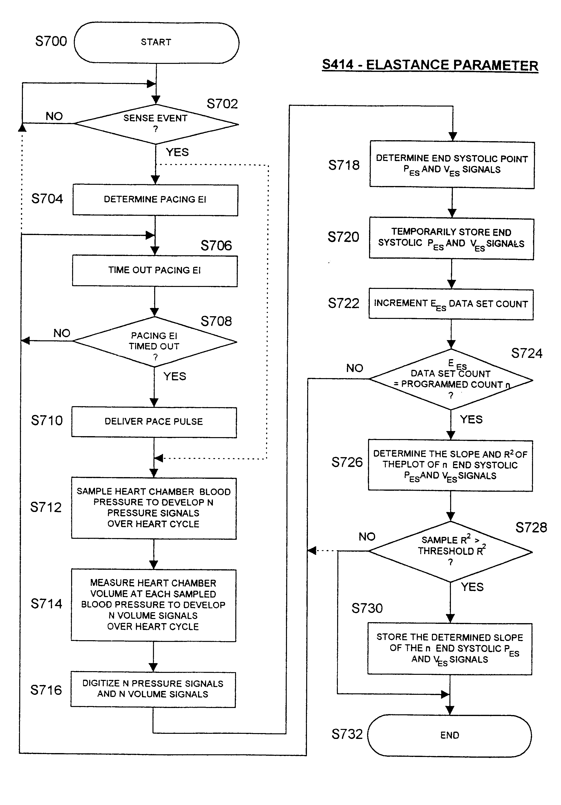
The implantable stimulator and monitor is operated in a one or more of the measurement modes that, in some instances, require delivery of an extrasystolic (ES) pulse after an extrasystolic interval (ESI) to induce PESP effects that are measured. In the present invention, the PESP capability is also employed to strengthen the cardiac contraction when one or more of the MR, RF, tau, and E.sub.ES parameters show that the heart condition has progressed to benefit from increased contractility, decreased relaxation time, and increased cardiac output. In this context, the stimulation therapy is referred to as PESP stimulation or pacing. In accordance with the invention, the effects of the applied PESP stimulation therapy can be observed over time by entering a heart function parameter measuring mode and gathering the parameter data.
The present invention combines these approaches, rendering a device that detects and monitors levels of cardiac function and delivers or modifies a therapy on the basis of this monitored information. The primary mode of delivery is direct electrical stimulation, resulting in improved contractility, relaxation, pressures or cardiac output.

Problems solved by technology

However, certain of the parameters are only measured or certain of the data are only stored when the patient heart rate is regular and within a normal sinus range between programmed lower and upper heart rates.

Method used


Image

  • Implantable medical device for treating cardiac mechanical dysfunction by electrical stimulation
  • Implantable medical device for treating cardiac mechanical dysfunction by electrical stimulation
  • Implantable medical device for treating cardiac mechanical dysfunction by electrical stimulation

Examples


Embodiment Construction

In the following detailed description, references are made to illustrative embodiments for carrying out the invention. It is understood that other embodiments may be utilized without departing from the scope of the invention.

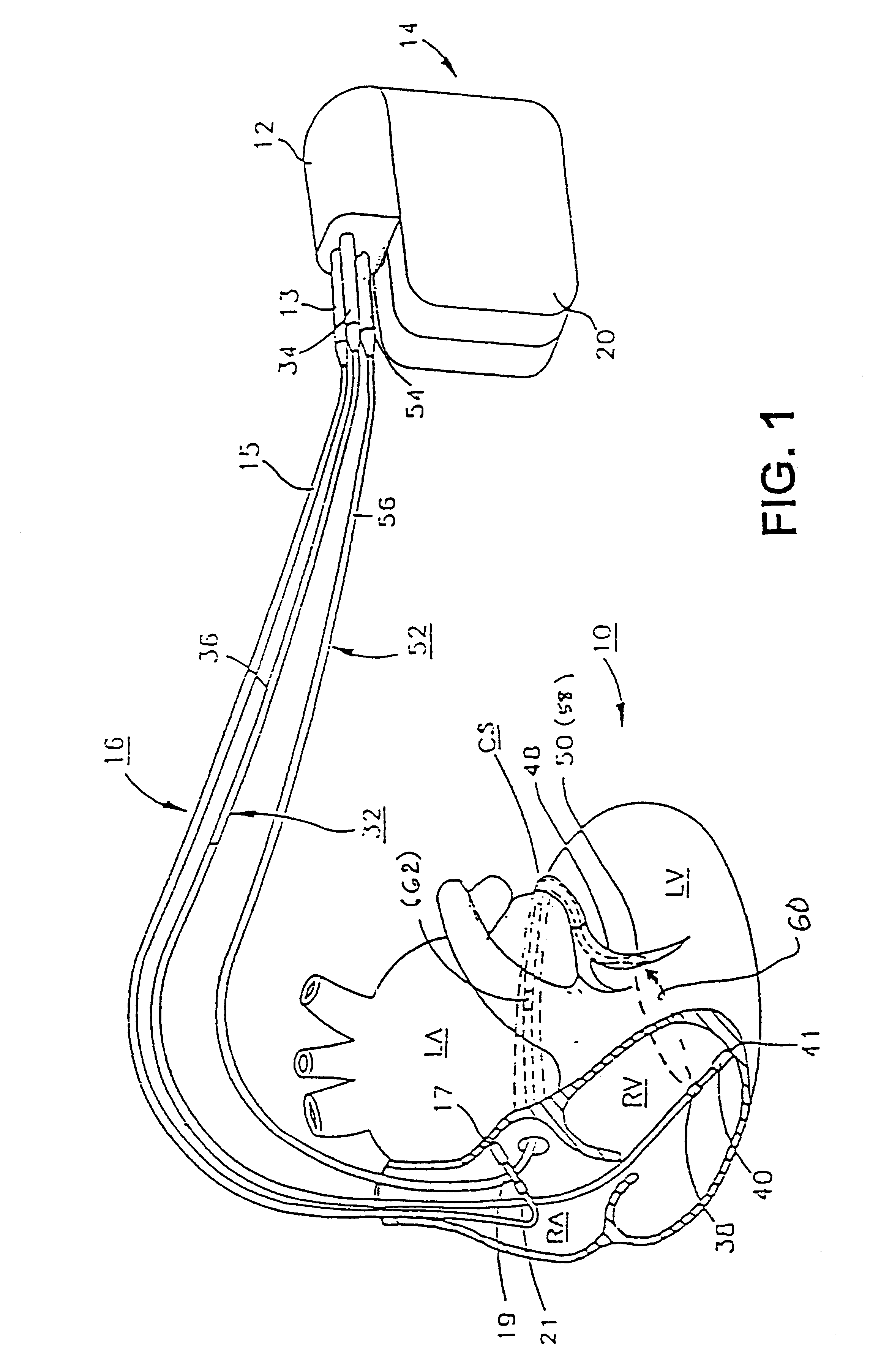
Before describing the preferred embodiments, reference is made to FIG. 22 reproduced from the above-referenced '464 patent which depicts the electrical depolarization waves attendant a normal sinus rhythm cardiac cycle in relation to the fluctuations in absolute blood pressure, aortic blood flow and ventricular volume in the left heart. The right atria and ventricles exhibit roughly similar pressure, flow and volume fluctuations, in relation to the PQRST complex, as the left atria and ventricles. It is understood that the monitoring and stimulation therapy aspects of this invention may reside and act on either or both sides of the heart. The cardiac cycle is completed in the interval between successive PQRST complexes and following relaxation of the atria and ve...


PUM


Abstract

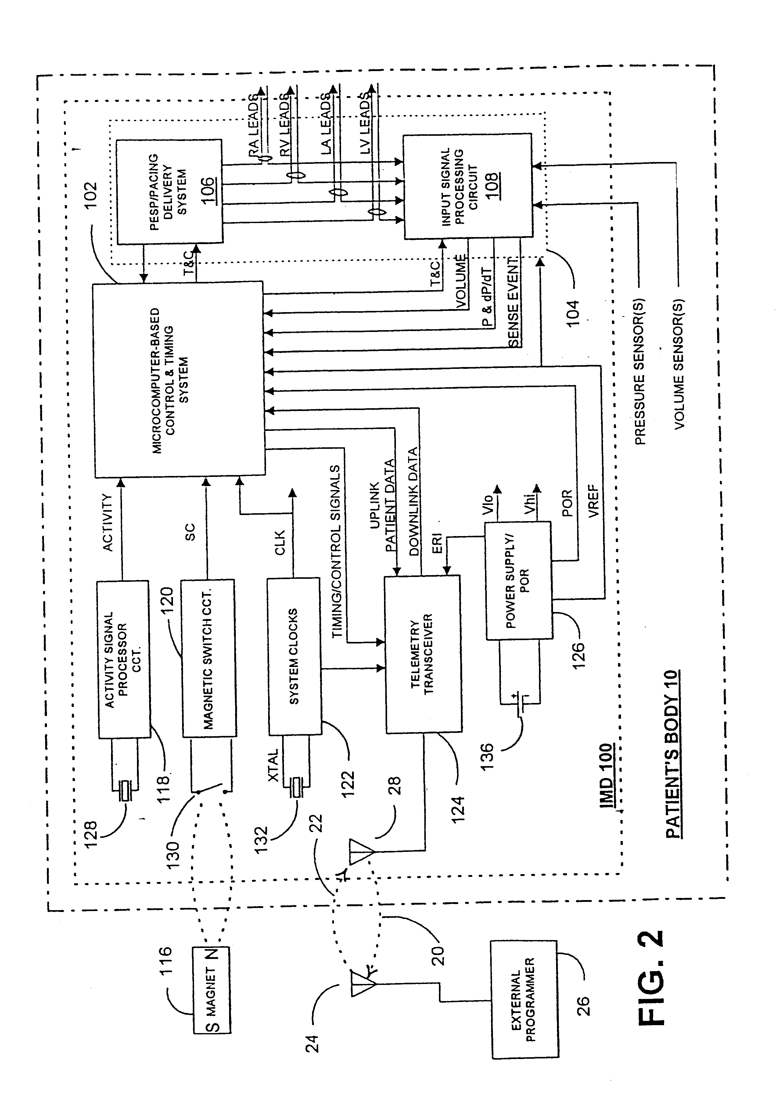
An implantable stimulator and monitor measures a group of heart failure parameters indicative of the state of heart failure employing EGM signals, measures of blood pressure including absolute pressure P, developed pressure (DP=systolic P-diastolic P), and / or dP / dt, and measures of heart chamber volume (V) over one or more cardiac cycles. These parameters include: (1) relaxation or contraction time constant tau (.tau.); (2) mechanical restitution (MR), i.e., the mechanical response of a heart chamber to premature stimuli applied to the heart chamber; (3) recirculation fraction (RF), i.e., the rate of decay of PESP effects over a series of heart cycles; and (4) end systolic elastance (E.sub.ES), i.e., the ratios of end systolic blood pressure P to volume V. These heart failure parameters are determined periodically regardless of patient posture and activity level. The physician can determine whether a particular therapy is appropriate, prescribe the therapy for a period of time while again accumulating the stored patient data for a later review and assessment to determine whether the applied therapy is beneficial or not, thereby enabling periodic changes in therapy, if appropriate. Drug therapies and electrical stimulation therapies, including PESP stimulation, and pacing therapies including single chamber, dual chamber and multi-chamber (bi-atrial and / or bi-ventricular) pacing can be delivered. In patient's prone to malignant tachyarrhythmias, the assessment of heart failure state can be taken into account in setting parameters of detection or classification of tachyarrhythmias and the therapies that are delivered.

Description

FIELD OF THE INVENTIONThe present invention relates generally to implantable medical devices and more specifically to monitoring signs of acute or chronic cardiac mechanical dysfunction such as congestive heart failure (CHF) or cardiogenic shock and providing appropriate therapies.BACKGROUND OF THE INVENTIONPatients suffering from chronic CHF manifest an elevation of left ventricular end-diastolic pressure, according to the well-known heterometric autoregulation principles espoused by Frank and Starling. This may occur while left ventricular end-diastolic volume remains normal due to a decrease in left ventricular compliance concomitant with increased ventricular wall stiffness. CHF due to chronic hypertension, ischemia, infarct or idiopathic cardiomyopathy is associated with compromised systolic and diastolic function involving decreased atrial and ventricular muscle compliance. These may be conditions associated with chronic disease processes or complications from cardiac surgery ...

Claims


Application Information

Patent Timeline
Patent Type & Authority Patents(United States)
IPC IPC(8): A61N1/365A61B5/05A61N1/362A61B5/0215
CPCA61N1/3627A61N1/36564
Inventor DENO, D. CURTISMULLIGAN, LAWRENCE J.BENNETT, TOM D.IGEL, DAVID A.HILL, MICHAEL R. S.SHAW, RICHARD J.
Owner MEDTRONIC INC
Learn More