Method of computed tomography using fluorinated gas-filled lipid microspheres as contrast agents

a computed tomography and lipid microsphere technology, applied in the direction of ultrasonic/sonic/infrasonic diagnostics, dispersion delivery, diagnostics, etc., can solve the problems of bubbles not encapsulated, no suggestion that these compositions would be useful as contrast media, and no suggestion that these liposomes could serve as contrast media for computed tomography

Inactive Publication Date: 2000-09-12
IMARX THERAPEUTICS
View PDF308 Cites 31 Cited by
  • Summary
  • Abstract
  • Description
  • Claims
  • Application Information

AI Technical Summary

Benefits of technology

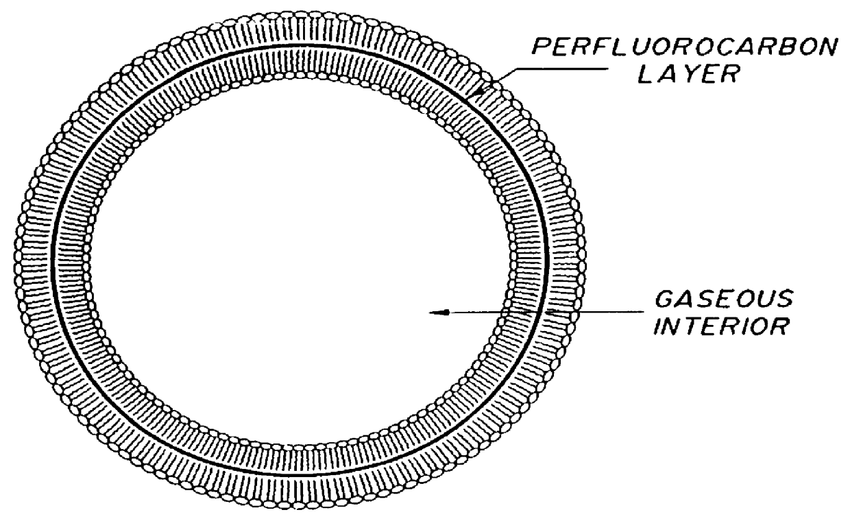
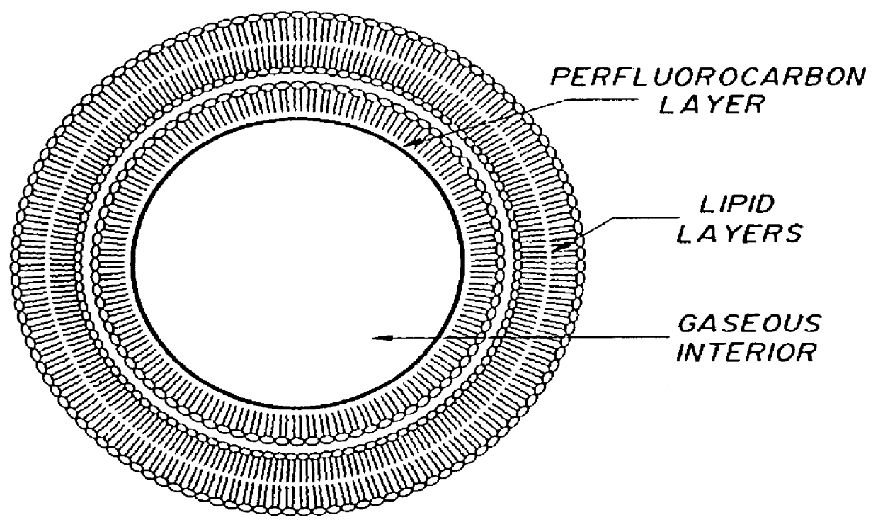
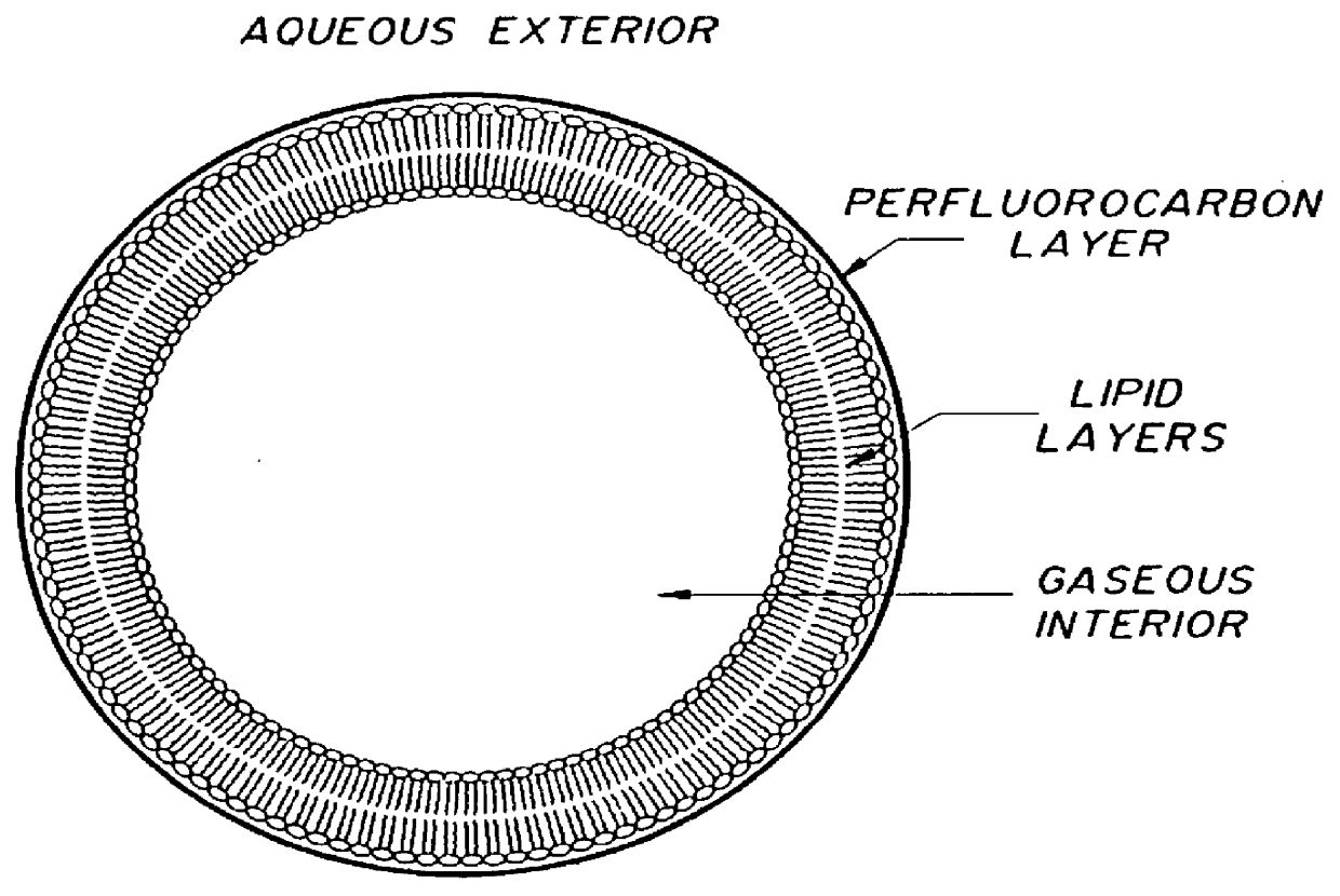
All of the above aspects of the present invention can be carried out, often with considerable attendant advantage, especially with regard to ease of ingestion by a patient, by using gaseous precursors to form the gas of the gas-filled microspheres. Once ingested, and upon gas formation in, for example, the gastrointestinal tract, expansion of the gaseous precursor causes an increase in the volume of the contrast medium and impart low density to the gastrointestinal tract, thereby enhancing computed tomography imaging thereof. These gaseous precursors may be activated by a number of factors, but preferably are temperature activated, that is, they are activated by exposure to elevated temperature. Such gaseous precursors are compounds which, at a selected activation or transition temperature, change phases from a liquid to a gas. Activation thus takes place by increasing the temperature of the compound from a point below, to a point above, the activation or transition temperature. Optionally, the contrast medium may further comprise a liquid fluorocarbon compound, for example, a perfluorocarbon, to further stabilize the microspheres. Preferably, the fluorocarbon liquid is encapsulated by the microspheres.

Problems solved by technology

However, there is no suggestion that these compositions would be useful as contrast media for computed tomography.
Again, however, these gas bubbles are not encapsulated and there is no suggestion of their use as contrast media for computed tomography.
However, there is no suggestion that these liposomes could serve as contrast media for computed tomography.
There is no suggestion, however, that these perfluorocarbon emulsions would be suitable for use as contrast media in computed tomography.
However, there is no suggestion that these liposomes can be gaseous precursor filled or that they might serve as contrast media for computed tomography.
While thick walled microspheres filled with air, nitrogen or other gases can be used as CT contrast agents, the thick walls of such microspheres raise the effective density of the contrast medium, which may in turn limit the effectiveness of the contrast medium.
Furthermore, thick walled microspheres may be relatively unpalatable for oral ingestion, or may be difficult to metabolize following intravenous injection.
However, it is contemplated that difficulty may be encountered in determining whether a particular compound is a basic or an auxiliary agent, since the functioning of the compound in question is generally determined empirically, or by the results produced with respect to producing stabilized microspheres.
For example, the simple combination of a biocompatible lipid and water or saline, when shaken, will often give a cloudy solution subsequent to autoclaving for sterilization.
Such a cloudy solution may function as a contrast agent, but is aesthetically objectionable and may imply instability in the form of undissolved or undispersed lipid particles.
Further, the shaking may occur manually or by machine.
Unhydrated compound, especially lipid, appears as amorphous clumps of non-uniform size and is undesirable.
Further, the shaking may occur manually or by machine.

Method used


Image

  • Method of computed tomography using fluorinated gas-filled lipid microspheres as contrast agents
  • Method of computed tomography using fluorinated gas-filled lipid microspheres as contrast agents
  • Method of computed tomography using fluorinated gas-filled lipid microspheres as contrast agents

Examples


example 1

This example describes the preparation of gas and gas precursor filled microspheres within the scope of the invention.

DPPC (77.5 mole %), DPPA (12.5 mole %), and DPPE-PEG 5000 (10 mole %) were introduced into a carrier solution of normal saline with glycerol (10 wt. %) and propylene glycol (10 wt. %). To this mixture was added perfluoropentane and a portion of the suspension (6 mL) was placed in a 18 mL glass vial and autoclaved for 15 minutes at 121.degree. C. The resulting translucent suspension was allowed to cool to room temperature. No appreciable foam could be seen was observed, but gentle shaking produced a few small bubbles at the top of the suspension. Shaking on a Wig-L-Bug.TM. (Crescent Dental Mfg. Corp., Lyons, Ill.) for 2 minutes resulted in a dense foam that substantially filled the entire volume of the vial.

Samples of the lipid / perfluoropentane (PFP) suspension, with and without shaking, were scanned by computed tomography using a Siemens DRH Somatom Ill. (Siemens, Is...

example 2

This example is directed to an analysis the effect of manual and mechanical shaking on microsphere size.

A lipid / PFP suspension was prepared as described in Example 1. A sample of the suspension was shaken at room temperature manually (much less vigorously than with the Wig-L-Bug mechanical shaker utilized in Example 1). Substantially no foam was produced, only a few bubbles at the top of the liquid layer. However, when the sample was warmed to body temperature, 37.degree. C., i.e., above the 27.5.degree. C. boiling point of the perfluoropentane, and shaken manually, foam readily appeared and filled the entire vial. When the foam produced by the Wig-L-Bug mechanical shaker at room temperature was compared to the foam produced manually at body temperature, it was noted that the microspheres produced by manual shaking were somewhat larger than the microspheres produced by the Wig-L-Bug mechanical shaker. The microspheres produced by manual shaking rose to the surface more quickly than ...

example 3

This example is directed to the formation of stabilized gas-filled microspheres comprising lipid bilayers with polyvinyl alcohol.

The effect of a polymer, namely, polyvinylalcohol, on the size of microspheres containing perfluorocarbons is illustrated in this example. Gas-filled microspheres comprising a lipid were prepared by the addition of 5 mg / mL of a suspension of DPPC:phosphatidic acid, and DPPE-PEG 5000 in a molar weight ratio of 82:8:10 in a vehicle containing 5% by weight of polyvinylalcohol (weight average M.W. 50,000, 99+% hydrolyzed) in normal saline. To this mixture was added 50 .mu.L of perfluoropentane. An identical suspension to the above described suspension was also prepared except that the vehicle was normal saline, glycerol, and propylene glycol in a ratio of 8:1:1, v:v:v (Spectrum Chemical Co., Gardena, Calif.). The suspensions were then autoclaved at 121.degree. C. for 21 minutes in a Barnstead / Thermolyne autoclave (Barnstead / Thermolyne, Rancho Dominguez, Calif)...


PUM


Abstract

Novel gas filled microspheres useful as computed tomography (CT) contrast agents. The microspheres are prepared from a gas and / or a gaseous precursor, and one or more stabilizing compounds.

Description

The present invention relates to compositions for computed tomography. More particularly, the present invention relates to compositions for computed tomography which comprise gas-filled microspheres.Computed tomography (CT) is a diagnostic imaging technique which measures, in its imaging process, the radiodensity of matter. Radiodensity of matter is typically expressed in Hounsefield Units (HU). Hounsefield Units are a measure of the relative absorption of computed tomography X-rays by matter and is directly proportional to electron density. Water has arbitrarily been assigned a value of 0 HU, air a value of -1000 HU, and dense cortical bone a value of 1000 HU.Various tissues in the body possess similar densities. Difficulty has been encountered in generating by CT visual images of tissues which possess similar densities and which are proximate each other. For example, it is difficult to generate separate CT images of the gastrointestinal (GI) tract and adjacent structures, includin...

Claims


Application Information

Patent Timeline
IPC IPC(8): A61K49/18A61K49/04A61K49/06
CPCA61K49/0419A61K49/1806
Inventor UNGER, EVAN C.
Owner IMARX THERAPEUTICS
Why Eureka
Try Eureka