Bioelectrical cancer diagnosis of margins of a freshly dissected cancerous tumor
a bioelectrical and cancer technology, applied in the field of cancer diagnosis, can solve the problems of at least 15-20% misdiagnosis, difficult accurate diagnosis of frozen sections, opaque hematoxylin-eosin staining and hence misdiagnosis, etc., to facilitate the placement of respective distal ends, facilitate the use of vacuum pressure, and facilitate the effect of utilizing th
- Summary
- Abstract
- Description
- Claims
- Application Information
AI Technical Summary
Benefits of technology
Problems solved by technology
Method used
Image
Examples
example 1
on of a Bioimpedance Sensor
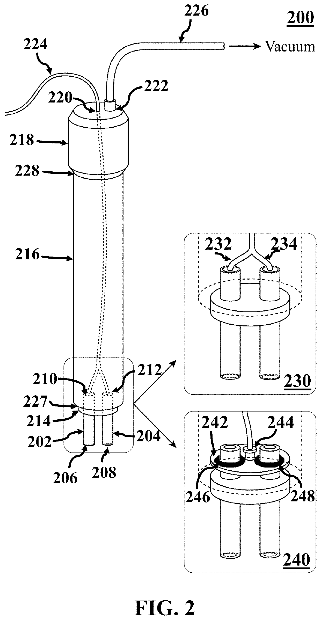
[0096]In this example, an exemplary bioimpedance sensor similar to bioimpedance sensor 102 was designed and fabricated. FIG. 6 shows an image of an exemplary head part of an exemplary fabricated bioimpedance sensor 600, consistent with one or more exemplary embodiments of the present disclosure. Exemplary fabricated bioimpedance sensor 600 includes two electrodes 602 and 604, which include two medical-grade stainless steel Veterinary Hypodermic G14 needles that were cut and polished to make 15 mm long needle tubes 602 and 604. An outer and an inner diameter of the needle tubes 602 and 604 are about 2 mm and about 1 mm, respectively. Needle tubes 602 and 604 were shielded by two plastic covers (with black color in FIG. 6), so that only the electrodes' cross surface may be in contact with a tumor tissue. Needle tubes 602 and 604 were embedded in exemplary electrode holder 606 with distance 610 of about 4 mm, and also needle tubes 602 and 604 were soldered to...
example 2
Cancerous Status of Margins of Dissected Tumors from Mice
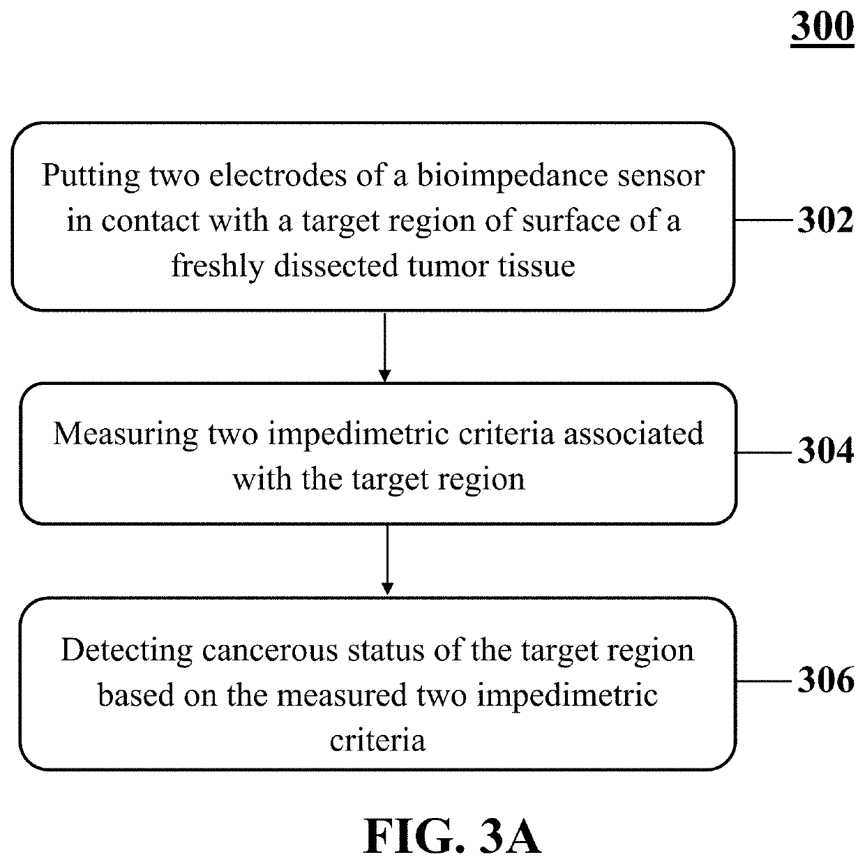
[0097]In this example, 10 female BALB / C mice that were 5 to 6 weeks old were tumorized with 4T1 cell line. 4T1 cell line is a mouse type breast cancer cell line with invasive phenotypes. 4T1 Cells were kept in DMEM culture medium complimented with 5% fetal bovine serum and 1% penicillin / streptomycin at 37° C. (5% CO2, 95% filtered air). A manual cell counting method (i.e., haemocytometer neubauer) was used to determine primary populations of the cultured cell lines. 10 female BALB / C mice were tumorized by subcutaneously implanting of about 2×10⁶ / 0.2 ml−1 4T1-derived cancer cells into back of the 10 female BALB / C mice under 50 mg / kg of ketamine and 10 mg / kg of xylazine anesthesia. The 10 female BALB / C mice were maintained in individual groups with similar size of formed tumors with sharp histological distinct patterns. After about 14 days, exemplary method 300 was applied to normal and tumoral regions of dissected cancerous mas...
example 3
Cancerous Status of Margins of Human Dissected Tumors
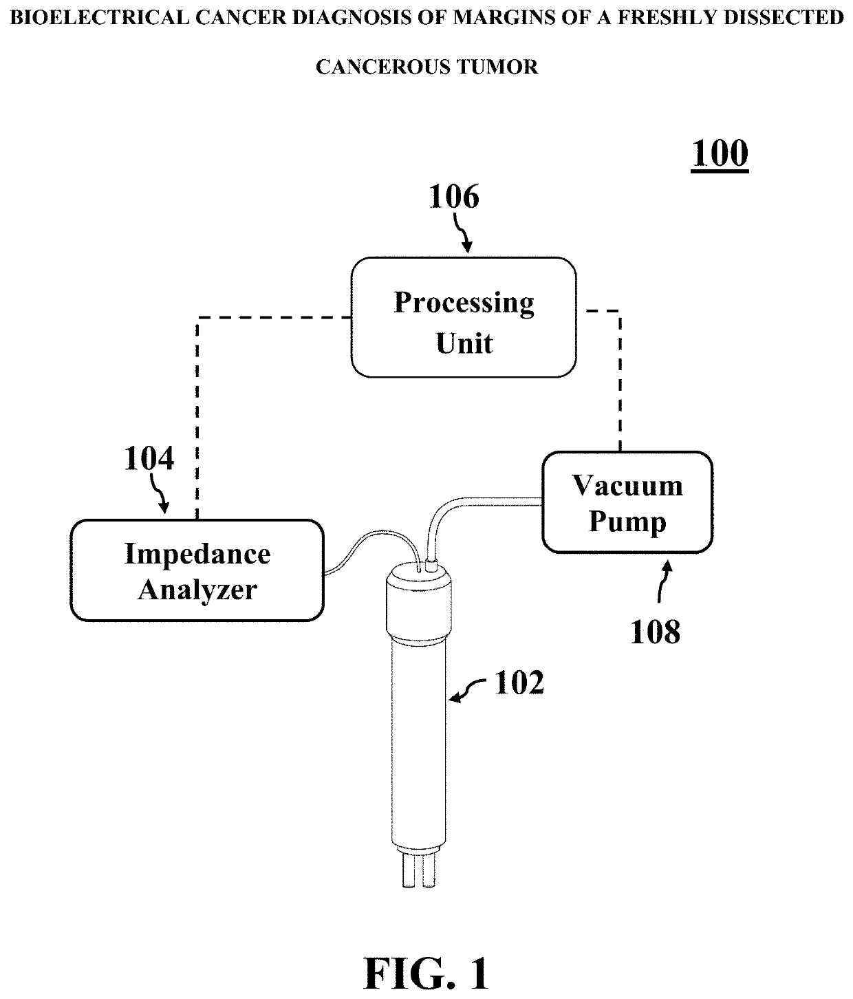
[0103]In this example, an exemplary method similar to method 300 utilizing exemplary fabricated bioimpedance sensor 600 and system 100 was applied to fresh margin tissues dissected from breast cancer patients which have been sent for intraoperative frozen-section. Impedance spectroscopy of 313 different samples (e.g., cancerous tumors, benign lesions, fibro-fatty tissues, neo-adjuvant mastectomy cases, etc.) obtained from surface of masses had been dissected from 68 patients accomplished in a frequency range of 1 Hz to 1 MHz.
[0104]After preparing touch imprint cytology slides from dissected breast tumor masses (first step of the intraoperative frozen-section in pathology labs), margins were pre-evaluated by a pathologist. Then, steps of exemplary method 300 was applied on tumor margins, before the main frozen-section process, to record an impedance spectroscopy of superficial surface all around an exemplary dissected breast tumor ...
PUM
 Abstract
Description
Claims
Application Information
 - R&D
- Intellectual Property
- Life Sciences
- Materials
- Tech Scout
- Unparalleled Data Quality
- Higher Quality Content
- 60% Fewer Hallucinations
Browse by: Latest US Patents, China's latest patents, Technical Efficacy Thesaurus, Application Domain, Technology Topic, Popular Technical Reports.
© 2025 PatSnap. All rights reserved.Legal|Privacy policy|Modern Slavery Act Transparency Statement|Sitemap|About US| Contact US: help@patsnap.com


