Method and device for in situ cancer margin detection

Active Publication Date: 2019-04-25
PURDUE RES FOUND INC
View PDF0 Cites 1 Cited by
  • Summary
  • Abstract
  • Description
  • Claims
  • Application Information

AI Technical Summary

Benefits of technology

This patent describes a system and method for detecting tumor borders using ultrasound and photoacoustic imaging. The system includes an imaging and control console with ultrasound and data acquisition subsystems. The laser subsystem includes a laser and a quartz crystal for adjusting optical energy. The imaging probe has an ultrasound transducer and an optical channel for delivering optical energy to the tissue. The system can also have a scanning stage for holding the tissue or imaging probe. The method involves fixing the tissue in a cartridge, scanning one side with ultrasound and photoacoustic signals, and then repositioning the focus spot for a second scan. The resulting images can be combined to create a 3D image. The system can also identify specific tissue types based on the ultrasound and photoacoustic images.

Problems solved by technology

Such confirmation is usually done by post-surgical histopathology or intraoperative pathology, which are time consuming and labor intensive.
It is a challenge for most surgeries of solid tumors, such as breast cancer, brain cancer, skin cancer, kidney cancer, and liver cancer etc.
Cytological examination and frozen section are widely applied clinically, but these two methods suffer from long procedure time and low sensitivity owning to the sampling method.
Radio frequency spectroscopy reduces the procedure time, but suffers from the sensitivity and specificity due to the lack of chemical selectivity.
Intraoperative ultrasound imaging has been applied to guide lumpectomies but with poor chemical selectivity.
Emerging optical technologies, including diffuse reflectance imaging, optical coherence tomography and spatial offset Raman spectroscopy, have greatly improved the sensitivity and specificity but still suffer from long procedure time, shallow tissue penetration, or an inability to assess the entire tumor tissue.
This technique requires exogenous labels to specific cancer targets, raising issues of labeling efficiency, toxicity, and regulatory burden.
However, it suffers from poor depth resolution due to a dramatic decrease in depth sensitivity.

Method used


Image

  • Method and device for in situ cancer margin detection
  • Method and device for in situ cancer margin detection
  • Method and device for in situ cancer margin detection

Examples


Embodiment Construction

[0034]For the purposes of promoting an understanding of the principles of the present disclosure, reference will now be made to the embodiments illustrated in the drawings, and specific language will be used to describe the same. It will nevertheless be understood that no limitation of the scope of this disclosure is thereby intended.

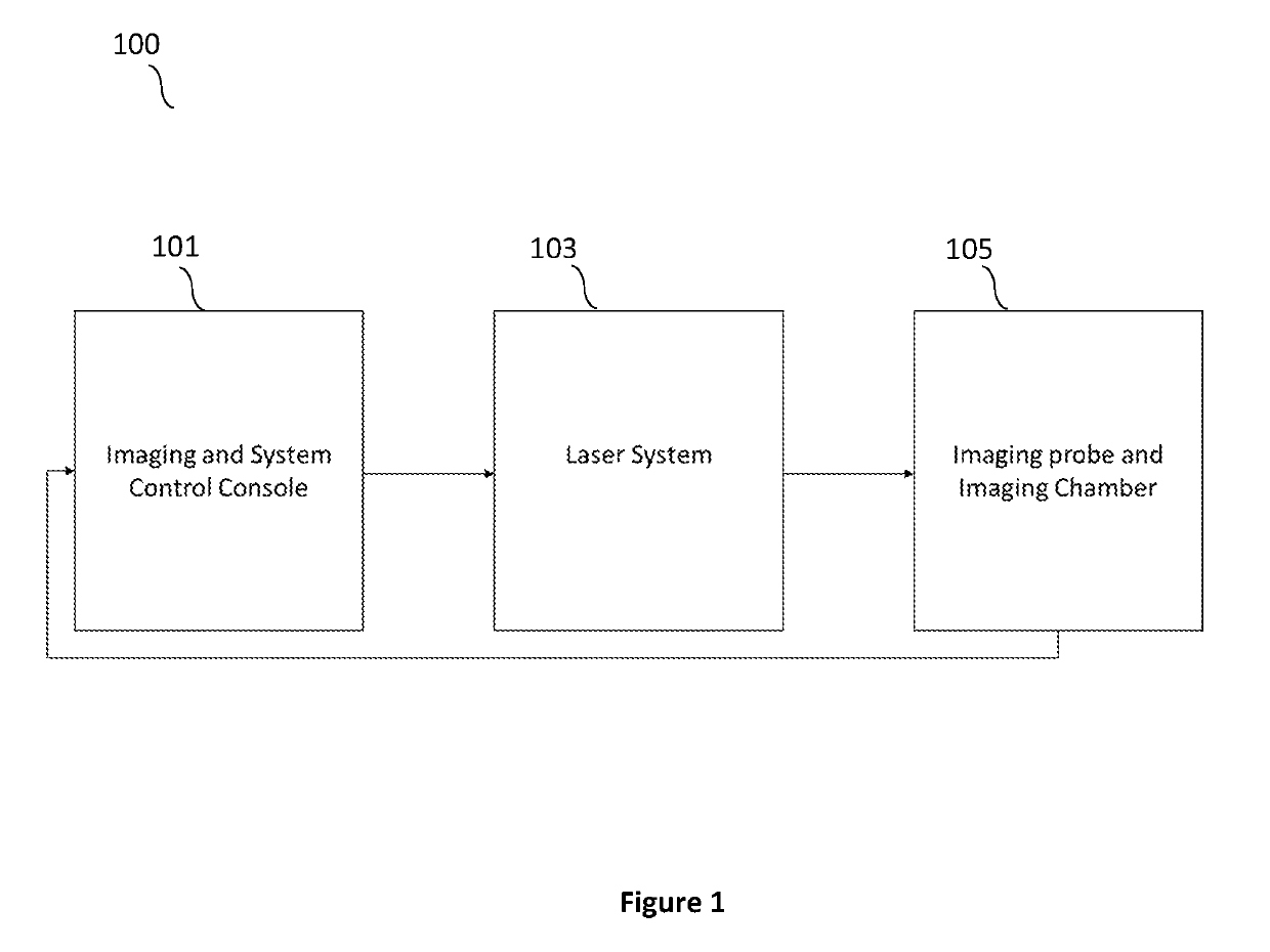
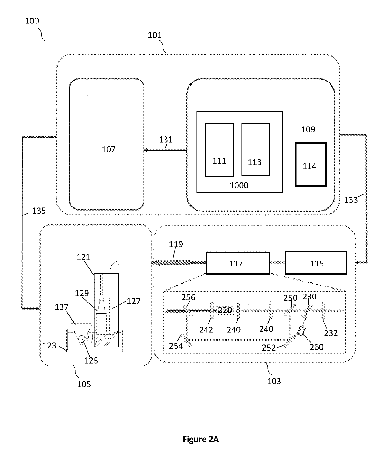
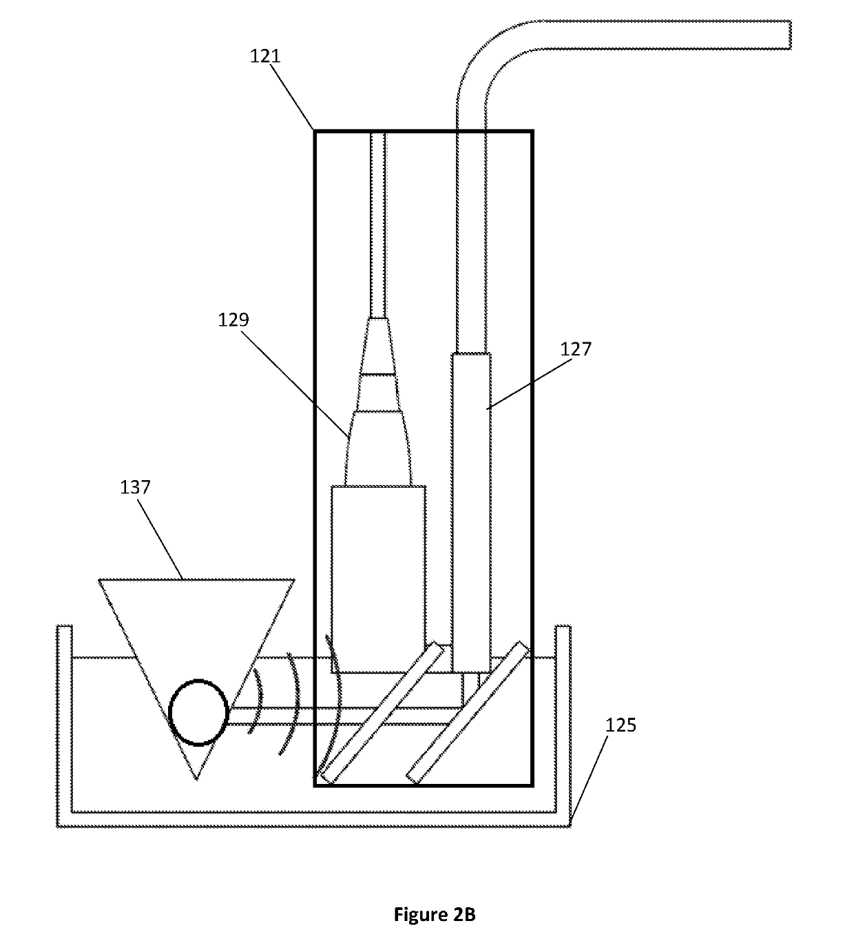
[0035]A novel arrangement that can offer a high-speed and highly-sensitive intraoperative assessment of breast cancer margin during conservation surgical procedures is disclosed herein. In particular, a multi-modal ultrasound / photoacoustic (US / PA) imaging system for highly sensitive breast cancer margin assessment is presented. Photoacoustic tomography (PAT) has proved its capability of rapid deep tissue imaging with chemical selectivity. It also incorporates an ultrasound imaging modality. With hemoglobin, adipose tissue, and photoacoustic contrast agents as the contrast, the PAT of the present disclosure can be applied in breast cancer imaging for sur...


PUM


Abstract

A multimodal ultrasound / photoacoustic tumor margin detection system. The system can be comprised of an imaging and system control console, which can include an ultrasound and data acquisition subsystem including an ultrasound transmitter and receiver configured to transmit and receive ultrasonic energy. A host-control computer configured to provide a function generator function and a delay generation function can also be a part of the imaging and system control console.

Description

CROSS-REFERENCE TO RELATED APPLICATION[0001]This U.S. Patent Application claims priority to U.S. Provisional Application: 62 / 249,747 filed Nov. 2, 2015, and U.S. Provisional Application: 62 / 295,028 filed Feb. 13, 2016 the disclosures of which is considered part of the disclosure of this application and is hereby incorporated by reference in its entirety.FIELD OF THE INVENTION[0002]The present application relates to optical detection systems and in particular to a photoacoustic detection system that can be used in surgical procedures.BACKGROUND[0003]This section introduces aspects that may help facilitate a better understanding of the disclosure. Accordingly, these statements are to be read in this light and are not to be understood as admissions about what is or is not prior art.[0004]As with all cancers, early diagnosis and treatment including surgical excision are chief amongst improving survival chances. Confirmation of the tumor removal during cancer surgery is critical for lowe...

Claims


Application Information

Patent Timeline
IPC IPC(8): A61B8/08A61B5/00A61B8/00
CPCA61B8/5223A61B5/0095A61B8/4281A61B2505/05A61B2576/02A61B8/0825A61B8/085A61B8/4416G16H50/30
Inventor CHENG, JI-XINWANG, PULAN, LULI, RUI
Owner PURDUE RES FOUND INC
Why Eureka
Try Eureka