Apparatus and method for assessment of cancer margin

a cancer margin and applicator technology, applied in the field of applicator and method for the assessment of cancer margins, can solve the problems of long-term detrimental effects on patient outcome, limited surgical techniques for resecting cancer, and excessive removal of healthy tissu

Inactive Publication Date: 2019-08-29
KERSEY ALAN D
View PDF13 Cites 1 Cited by
  • Summary
  • Abstract
  • Description
  • Claims
  • Application Information

AI Technical Summary

Benefits of technology

[0018]The present invention is concerned with an apparatus and method for inspecting a biological tissue using a pH-sensitive coating material to determine whether the tissue is normal or cancerous. The coating material is placed in contact to the tissue and excited by an excitation light. The coating material contains one or more pH sensitive dyes arranged to provide a response signal indicative of the pH value of the tissue in response to the excitation. Using a fiber bundle having a plurality of optical fibers formin

Problems solved by technology

Current surgical techniques to resect cancer are limited by the lack of a precise method to determine the boundary between normal and cancerous cells in, known as the ‘tumor margin’, in real time during surgical procedures.
As a result, surgeons often perform what is called cavity shaving, which can result in the removal of excessive amounts of healthy tissue.
Conversely, many patients do not have the entire tumor removed during the initial surgery and may need a follow up surgery to remove residual cancer tissue.
This can be traumatic to the cancer patient, adding stress, resulting in long-term detrimental effects on the patient outcome.
While these were used

Method used


Image

  • Apparatus and method for assessment of cancer margin
  • Apparatus and method for assessment of cancer margin
  • Apparatus and method for assessment of cancer margin

Examples


Embodiment Construction

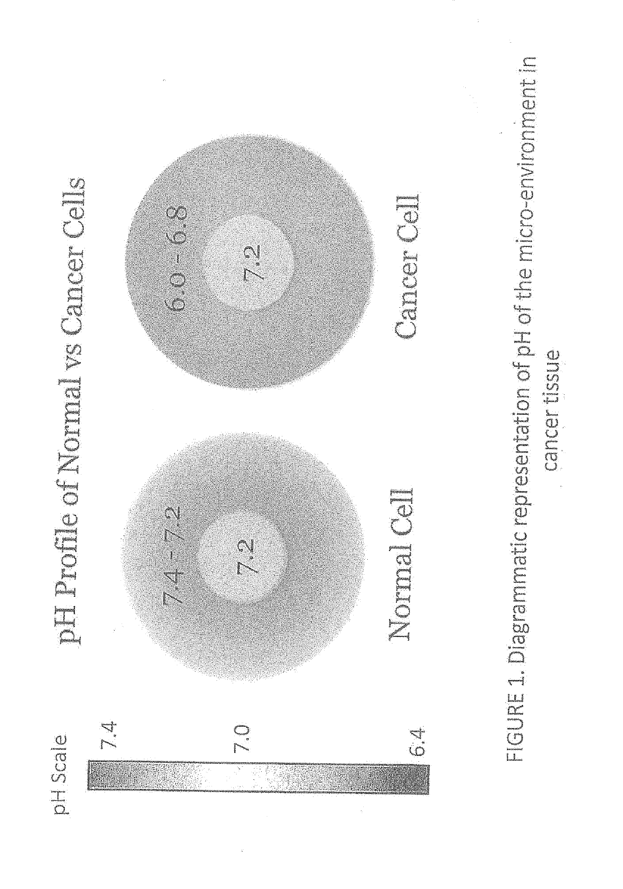
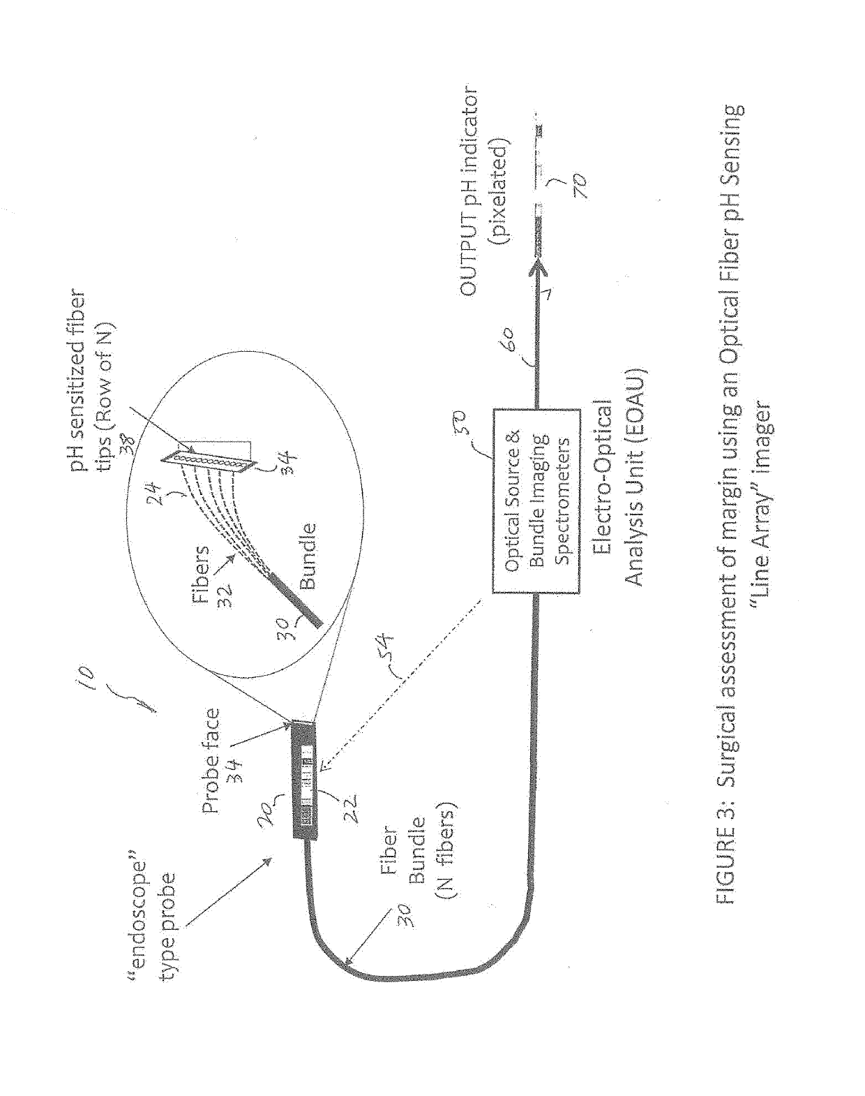
[0056]The present invention makes use of a bundled fiber-optic probe for the imaging of localized surface pH in a biological tissue and specifically for the determination of tumor margin or the boundary between normal and cancerous cells. The approach also provides direct mapping of the tumor margin via a display that could be provided for a surgeon's or pathologist's inspection, or via a display mapped back onto the probe via a second fiber optic bundle that allows the surgeon to see the pH map as soon as the probe is contacted with the tissue surface. FIGS. 3 to 7 show some representative examples of the present invention.

[0057]FIG. 3 illustrates an apparatus for inspecting a biological tissue, according to an embodiment of the present invention. The apparatus is illustrated as a ‘line array’ imager embodiment where fiber optic tip probes sensitized to local pH are arranged to allow visualization of the tumor margin, e. g., the transition from tumor to normal tissue. As an example...


PUM


Abstract

An apparatus for inspecting a biological tissue uses a pH-sensitive coating material to determine whether the tissue is normal or cancerous. The coating material is placed in contact with the tissue to be excited by an excitation light. The coating material is arranged to provide a response signal indicative of the pH value of the tissue. Using a fiber bundle having a plurality of optical fibers forming a linear array or a two-dimensional array adjacent the coating material, the imaging of localized surface pH in the biological tissue can be achieved using the response signal through each of the optical fibers. The fiber bundle can be arranged as a probe to examine the tissue for providing direct mapping of the tumor margin via a display, so that a surgeon can inspect the tissue in real-time.

Description

CROSS REFERENCE TO RELATED PATENT APPLICATION[0001]This application claims priority to U.S. Provisional Application No. 62 / 591,335, filed Nov. 28, 2017, whose entire content is hereby incorporated by reference.FIELD OF THE INVENTION[0002]The present invention relates generally to an apparatus and method for the assessment of cancer margin, and more specifically to the assessment during surgical procedures using a fiber optic pH sensing array with real-time visual mapping.BACKGROUND[0003]For many decades the reference method for the diagnosis of cancer has been histopathological examination of tissues using conventional microscopy. This process is known as Surgical Pathology. In Surgical Pathology, samples can be produced from surgical procedures (tumor resection), diagnostic biopsies or autopsies. These samples go through a process that includes dissection, fixation, and cutting of tissue into precisely thin slices which are stained for contrast and mounted onto glass slides. The sl...

Claims


Application Information

Patent Timeline
IPC IPC(8): A61B5/1459A61B1/00A61B5/145A61B5/00G01N33/574
CPCA61B5/1459A61B1/0017A61B5/14539A61B5/0084A61B5/0075G01J3/44A61B1/00167G01N21/65G01J2003/4424G01N21/274G01N33/574A61B5/14546G01N33/4833
Inventor KERSEY, ALAN D.
Owner KERSEY ALAN D
Why Eureka
Try Eureka