Anti-pd-1 nano-antibody and application thereof
a nano-antibody and anti-pd technology, applied in the field of biomedical or biopharmaceutical technology, can solve the problems of limited tumor penetration and slow distribution, and achieve the effect of blocking binding and good
- Summary
- Abstract
- Description
- Claims
- Application Information
AI Technical Summary
Benefits of technology
Problems solved by technology
Method used
Image
Examples
example 1
n and Purification of Human PD-1 Protein
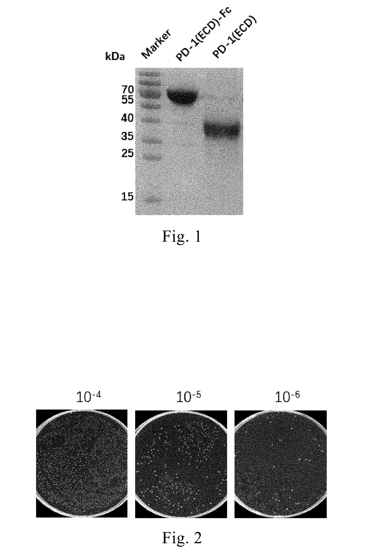
[0131](1) Firstly, the amino acid sequence and nucleotide sequence of human PD-1 were searched from UniProt, and the full-length nucleic acid sequence of PD-1 was integrated into pCDNA3.1(−) vector by the company, and the sequence of the extracellular domain was sub-cloned into pFUSE-IgG1 vector (commercially available from Invitrogen), wherein a TEV cleavage site and a GS linker were introduced at the end of the extracellular domain of PD-1 for later excision of the tag to obtain the Fc-free PD-1 (ECD) protein;
[0132](2) An Omega plasmid maxi kit was used to extract the constructed pFUSE-PD-1(ECD)-hIgG1-Fc2 plasmid;
[0133](3) HEK293F cells were cultured until an OD reached 2.0×10⁶ cells / mL;
[0134](4) The plasmid and the transfection agent PEI were mixed (1:3) well and placed in transfection medium F17 (Gibco) for 20 min, and then the product was added into HEK293F cells culture for further incubation in a shaker under 6% CO2 at 37° C. for 6 days...
example 2
ruction of PD-1 Nanobody Library
[0140](1) 1 mg of PD-1 (ECD) antigen was mixed with Freund's adjuvant in equal volume to immunize a Xinjiang bactrian camel once a week for a total of 7 times to stimulate B cells to express antigen-specific PD-1(ECD) nanobodies;
[0141](2) After the 7 immunizations were completed, 100 mL of camel peripheral blood lymphocytes were sampled and total RNA was extracted.
[0142](3) cDNA was synthesized and VHH was amplified using reverse PCR;
[0143](4) Phage display vector and VHH were digested with restriction endonucleases PstI and NotI, and the enzyme-cut VHH fragments were then ligated into the vector at a certain molar ratio at 16° C. for 16 h.
[0144](5) The ligated product was electronically transfected into competent TG1 cells, and the phage display library of the PD-1 nanobody was constructed and the capacity thereof was determined.
[0145]After detection, the library had a storage capacity of 2.3×10⁹ CFU (as shown in FIG. 2), and the correct insertion ra...
example 3
and Verification of PD-1 Nanobodies
[0146]Screening of Nanobodies
[0147](1) 10 μg PD-1 (ECD) antigens dissolved in 100 mM NaHCO3(pH 8.2) was added onto the NUNC ELISA plate and left overnight at 4° C. for coating;
[0148](2) The next morning, the antigen solution in the well was aspirated, washed with PBST (0.05% PBS+Tween-20) for 5 times, then 100 μl of 0.1% BSA was added, and blocked at room temperature for 2 h;
[0149](3) After 2 h, phages (2×10″ CFM of phage display gene library with nanobodies of immunized camel) were added and reacted at room temperature for 1 h;
[0150](4) Wash 5 times with PBST, to wash off phage display nanobodies that did not bind to antigen PD-1 (ECD);
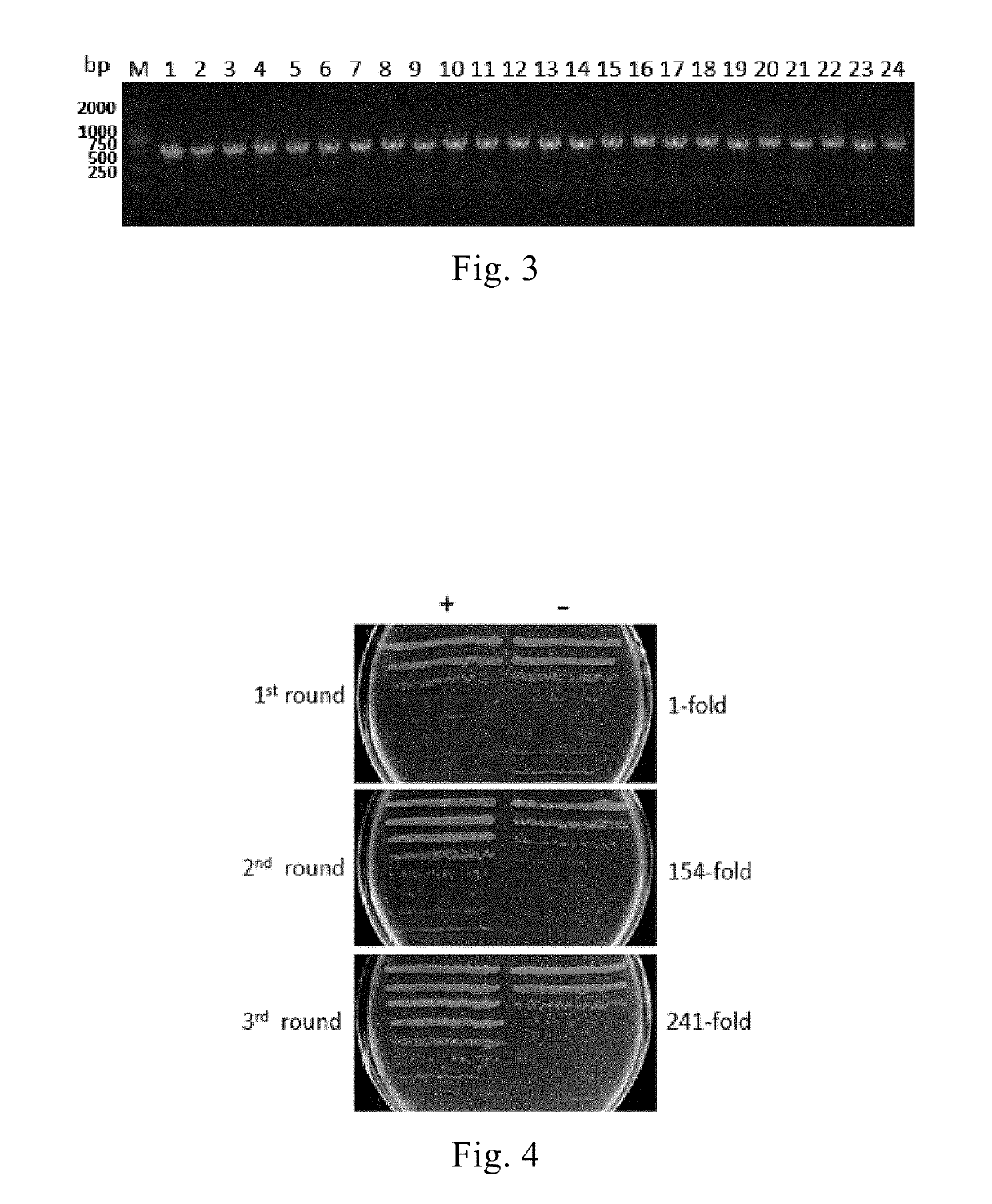
[0151](5) The phages specifically bound to PD-1 were dissociated by 100 mM triethanolamine, and E. coli TG1 cells in logarithmic phase were infected and incubated at 37° C. for 1 h. The phages were generated and purified for the next round of screening. The screening process was repeated for 3 rounds. The enrichment...
PUM
| Property | Measurement | Unit |
|---|---|---|
| temperature | aaaaa | aaaaa |
| pH | aaaaa | aaaaa |
| temperature | aaaaa | aaaaa |
Abstract
Description
Claims
Application Information
 - R&D
- Intellectual Property
- Life Sciences
- Materials
- Tech Scout
- Unparalleled Data Quality
- Higher Quality Content
- 60% Fewer Hallucinations
Browse by: Latest US Patents, China's latest patents, Technical Efficacy Thesaurus, Application Domain, Technology Topic, Popular Technical Reports.
© 2025 PatSnap. All rights reserved.Legal|Privacy policy|Modern Slavery Act Transparency Statement|Sitemap|About US| Contact US: help@patsnap.com


