Antidote for Chondroitinase

a chondroitinase and antidote technology, applied in the field of antidote for chondroitinase, can solve the problems of inactivation of chemical treatment enzymes, loss of hydration and function of discs, and spondyloepiphyseal dysplasia and premature osteoarthritis, and achieve the effect of inhibiting chondroitinase activity

Inactive Publication Date: 2019-08-15
WONG BING LOU
View PDF0 Cites 0 Cited by
  • Summary
  • Abstract
  • Description
  • Claims
  • Application Information

AI Technical Summary

Benefits of technology

[0013]The present invention provides a method for inhibiting chondroitinase activity in a substrate comprising introducing a non-substrate glycosaminoglycans (GAG) to the substrate containing the chondroitinase, wherein said non-substrate GAG is undegradable by the chondroitinase. The non-substrate glycosaminoglycans may be administered as a single dose or in multiple doses over a period of time. The chondroitinase may be Chondroitinase B, Chondroitinase C, Chondroitinase AC, and Chondroitinase ABC. The substrate may be one or more of dermatan sulfate, hyaluronic acid, chondroitin sulfate, or derivatives thereof. The non-substrate GAG may be from naturally unbranched homo-polysaccharide, unnaturally branched GAG, or a hybrid GAG m

Problems solved by technology

This CSPG is a critical structural element in cartilaginous tissue, and the loss of function of this molecule will result in spondyloepiphyseal dysplasia and premature osteoarthritis.
This is caused by gradually degradation of the chondroitin sulfate chain in the aggrecan molecule, resulting in loss of hydration and function of the disc.
The escaped NP together with the ruptured annulus fibrosus will compress the nearby nerve root, causing low back pain and sciatica.
For example, when using chondroitinase as chemonucleolysis agent for digestion of the nucleus pulposus, accidental over-dosing of the enzyme could happen during the operation or, alternatively, a patient coul

Method used


Image

  • Antidote for Chondroitinase
  • Antidote for Chondroitinase
  • Antidote for Chondroitinase

Examples


example 1

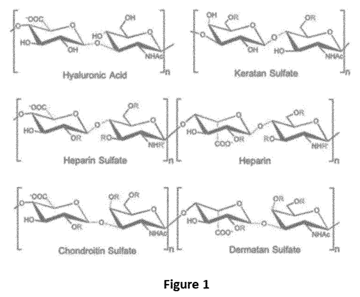
[0037]A Chondroitinase ABC model was generated from the crystal structure previously published (Huang W, Lunin V, Li Y, Suzuki S, Sugiura N, Miyazono H, Cygler M, Journal of Molecular Biology, (2003) 328, 623-634). A model of heparin tetrasaccharide was extracted and modified from the crystal structure of heparin (Khan, S, Gor, J, Mulloy, B, Perkins, S J, (2010) Journal of Molecular Biology 395: 504-521). The docking of the heparin tetrasaccharide to the chondroitinase ABC model was performed using Autodock v4.2. The heparin tetrasaccharide was docked within a grid box covering the whole active site with a size of 90×84×78 points using a spacing of 0.375 A. The parameters of the docking consisted of 30 Lamarckian Genetic Algorithm runs using 2,500,000 energy evaluations and a population size of 150 individuals. The docking result showed that the heparin tetrasaccharide had intensive interaction to the active residue of chondroitinase ABC, including His 501, Tyr 508, Arg 560, and His...

example 2

[0038]2 mg / ml Chondroitin sulfate C solution was prepared by dissolving in buffer at pH 8.0 Tris-HCl (50 mM). Freeze-dried chondroitinase ABC was reconstituted in 1 mg / ml BSA solution and was serially diluted to 0.5 U / ml by 1 mg / ml BSA. Both solutions were incubated at 37° C. for 5 min separately and were then mixed in a 3.5 ml quartz cuvette. The final concentration of chondroitinase ABC become 0.01 U / ml and 0.1 mg / ml chondroitin sulfate C. The reaction mixture was further incubated at 37° C. for 20 min in spectrophotometer and the absorbance at 232 nm was continuously monitored. The experiment was repeated in the presence of 0.0125 mg / ml heparin. In FIG. 3, the absorbance of the solution without the presence of heparin at 232 nm increased linearly. However, in the presence of 0.0125 mg / ml heparin, the enzymatic reaction of chondroitinase was completely inhibited.

example 3

[0039]EMSA was conducted by mixing 20 μg chondroitinase ABC (110 kDa, about 0.2 nmol) with 3.8 μg heparin (17-19kDa, about 0.2 nmol) at room temperature for 30 min. Each reaction mixture (16 μl), as well as samples with heparin and chondroitinase ABC, were mixed with 4 μl of 5× native loading buffer. The samples were loaded onto two separate 6% native gels for electrophoresis at 100V for 2.5 hours. After the electrophoresis, one gel was stained with Coomassie blue the other was stained by Alcian Blue. The gels are shown in FIG. 4 and FIG. 5. It can be seen in the gel stained with Alcian Blue (FIG. 4) that heparin is completely moved to the bottom of the gel in the absence of chondroitinase ABC. When Chondroitinase ABC is present, there is a significant amount of GAG present in the band that overlaps with the band of chondroitinase ABC. In other words, heparin is being retained by chondroitinase ABC, indicating that heparin binds tightly to the enzyme.


PUM


Abstract

The present invention provides a method for inhibiting chondroitinase activity in a substrate by introducing a non-substrate glycosaminoglycan (GAG) to the substrate containing the chondroitinase. The non-substrate GAG is undegradable by chondroitinase. The GAG may be administered as a single dose or in multiple doses. The chondroitinase may be Chondroitinase B, Chondroitinase C, Chondroitinase AC, or Chondroitinase ABC. The substrate may be one or more of dermatan sulfate, hyaluronic acid, chondroitin sulfate, or derivatives thereof. The non-substrate GAG may be from naturally unbranched homo-polysaccharide, unnaturally branched GAG, or a hybrid GAG molecule fused of two or three GAG chains, being produced by chemical synthesis or enzymatic reaction. The non-substrate GAG may be heparin, heparan sulfate, and keratan sulfate. The non-substrate GAG may bind to the active residue of the chondroitinase when being introduced to the substrate such that the chondroitinase is no longer enzymatically active.

Description

CROSS-REFERENCE TO RELATED APPLICATION[0001]The present application claims priority from the U.S. provisional patent application Ser. No. 62 / 630,810 filed Feb. 14, 2018, the disclosure of which is incorporated herein by reference in its entirety.TECHNICAL FIELD[0002]The present invention relates to a use of a non-substrate glycosaminoglycan (GAG)-based antidote for inhibiting chondroitinase activity in a substrate.BACKGROUND[0003]Glycosaminoglycan lyase (GAG lyase), which is a group of enzymes effectively degrading glycosaminoglycan (GAG), have been identified from viruses and a wide range of bacteria. In these micro-organisms, the enzymes are known to be used as a virulence factor for infection or as a digestive enzyme for utilizing GAG as carbon source. The substrates of the enzyme, GAGs, are negatively charged polysaccharides and are the major composition of extracellular matrix (ECM) synthesized in animals. Based on the chemical structure, GAG can be classified into 6 groups: ch...

Claims


Application Information

Patent Timeline
IPC IPC(8): C12N9/99
CPCC12N9/99
Inventor WONG, BING LOU
Owner WONG BING LOU
Why Eureka
Try Eureka