Optical coherence tomography for cancer screening and triage

Active Publication Date: 2019-07-25
RGT UNIV OF CALIFORNIA
View PDF0 Cites 9 Cited by
  • Summary
  • Abstract
  • Description
  • Claims
  • Application Information

AI Technical Summary

Benefits of technology

The present invention is an optical coherence tomography (OCT) imaging system integrated with an imaging probe that can provide better understanding of tissue substructure in real-time to screen for cancerous tissue. The system uses a continuously learning convolutional neural network (CNN) to make rapid classifications, which can guide a physician during an examination. The system includes a means for observing the sample region being scanned at the time of each imaging frame, which allows for precise co-registration of the gold standard histopathology and the imaging frame. The system also uses gradient refractive index (GRIN) rod lenses for imaging, which can cause unwanted spherical and chromatic aberrations. To account for such dispersion, image correction techniques, such as flattening, are used to reduce the impact of aberrations and increase processing speeds. The invention advantageously provides a real-time, low-cost, and non-invasive imaging system.

Problems solved by technology

Using GRIN rod lenses in the OCT imaging systems typically causes unwanted spherical and / or chromatic aberrations.
In addition, the use of a long piece of glass such as the GRIN rod will cause dispersion between the probe sample arm and reference arm in the OCT system.
This will result in a widening of the point spread function in the OCT signal, effectively reducing the resolution.
This is an inconvenience in optical design.

Method used


Image

  • Optical coherence tomography for cancer screening and triage
  • Optical coherence tomography for cancer screening and triage
  • Optical coherence tomography for cancer screening and triage

Examples


example 1

[0158]The following is a non-limiting example of the present invention. It is to be understood that said example is not intended to limit the present invention in any way. Equivalents or substitutes are within the scope of the present invention.

Introduction

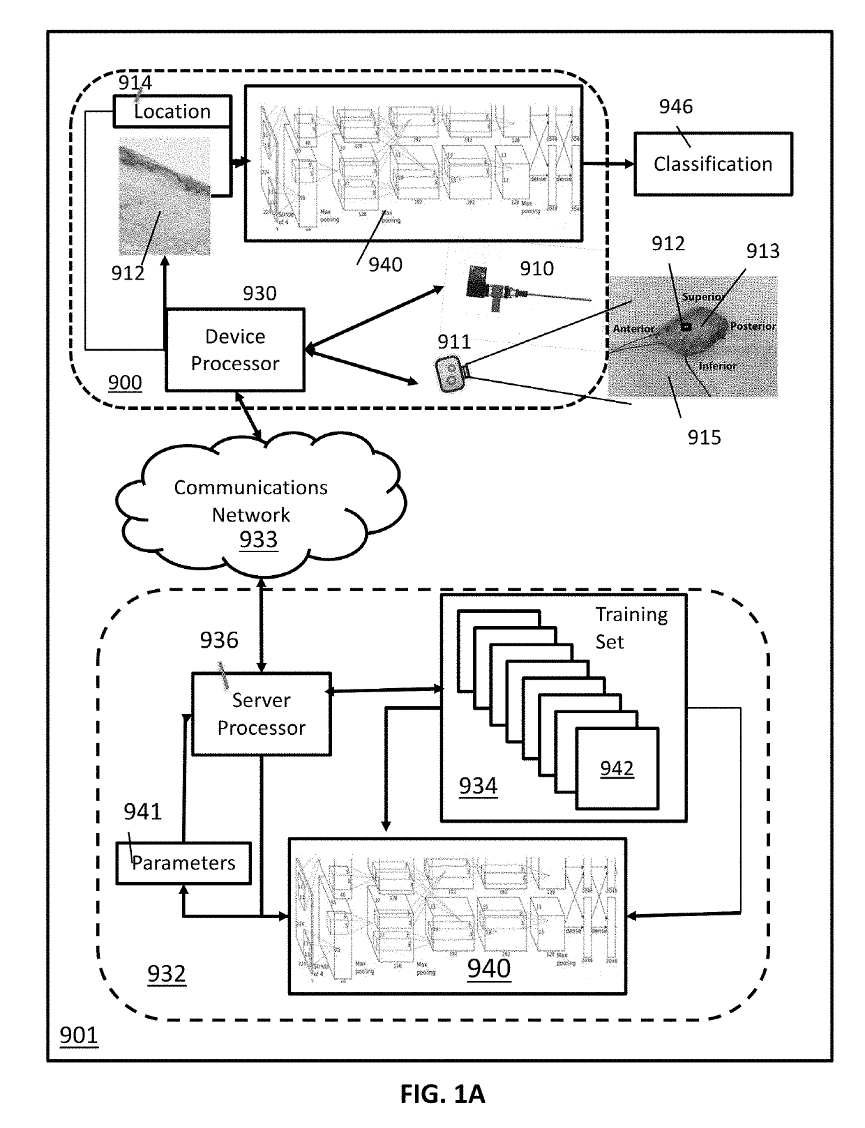
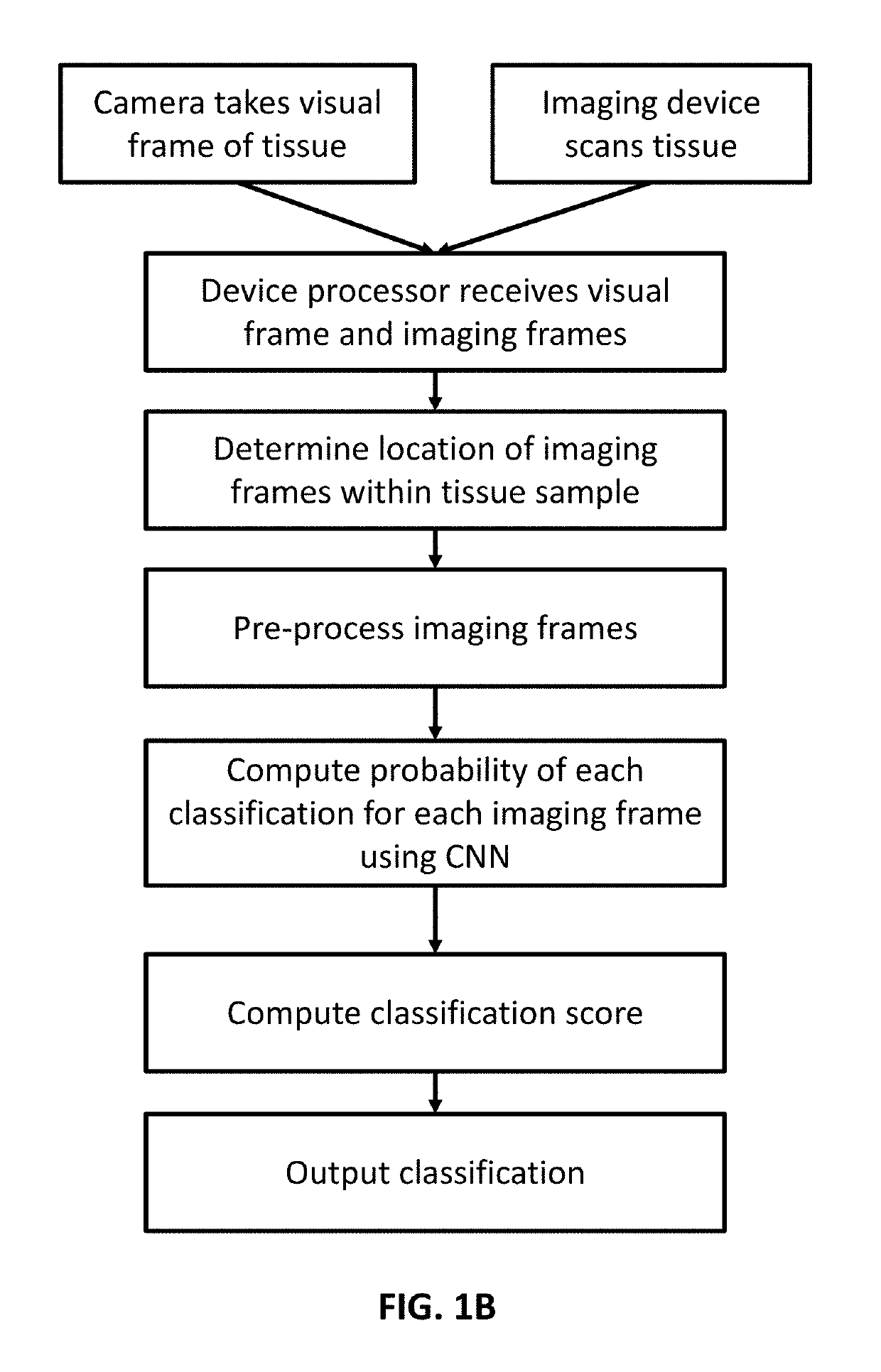
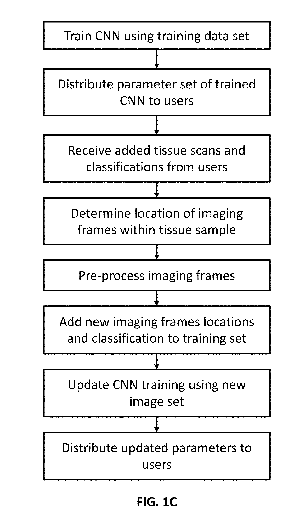
[0159]Incomplete surgical resection of head and neck cancer lesions is the most common cause of local cancer recurrence. Currently, surgeons rely on their experience of direct visualization, palpation, and pre-operative imaging to determine the extent of tissue resection. Intraoperative frozen section microscopy is used to assess presence of cancer at the surgical margin. It has been demonstrated that optical coherence tomography (OCT), a minimally invasive, non-ionizing near infrared mesoscopic imaging modality can resolve subsurface differences between normal and abnormal oral mucosa. However, previous work has utilized 2-D OCT imaging which is limited to the evaluation of a small regions of interest generated frame by frame. OC...


PUM


Abstract

A device for cancer screening and triage using an optical coherence tomography (OCT) imaging system integrated with optical imaging probe is provided. Endoscopic OCT images are generated using the OCT system having a helical probe. The images are further analyzed to generate a depth resolved intensity OCT signal to classify the region of tissue into variable grades of dysplasia to guide the physician's biopsy or resection.

Description

CROSS-REFERENCES TO RELATED APPLICATIONS[0001]This application is a non-provisional and claims benefit of Provisional U.S. Patent Application No. 62 / 619,537, filed Jan. 19, 2018, the specifications of which are incorporated herein in their entirety by reference.FIELD OF THE INVENTION[0002]The present invention relates to a device and a method for cancer screening and triage using an optical coherence tomography imaging system integrated with a side viewing fiber-optic probe and / or a forward viewing optical probe.BACKGROUND OF THE INVENTION[0003]Screening is an effective way to identify early stage cancer, such as oral squamous cell carcinoma (OSCC), and pulmonary cancer, to reduce its morbidity and mortality, for example. Cancer screening procedures that are fast, reliable, and less invasive are desired for improving diagnostic capabilities. As an example, oral squamous cell carcinoma (OSCC) ranks as the sixth most common cancer worldwide, accounting for approximately 400,000 new ca...

Claims


Application Information

Patent Timeline
IPC IPC(8): A61B5/00G01B9/02G06T7/50G06T7/13G06V10/764
CPCA61B5/0066G01B9/0205G01B9/02091G06T7/50G06T7/13A61B5/7267A61B5/0088A61B5/4552G06T7/0012G06T2207/30108G06T2207/10068G16H50/70G06T2207/20084G06V2201/03G06V10/82G06V10/764
Inventor HEIDARI, ANDREW EMONCHEN, ZHONGPINGBENN, BRYAN S.
Owner RGT UNIV OF CALIFORNIA
Why Eureka
Try Eureka