
Methods for repairing cartilage damage

a cartilage damage and cartilage technology, applied in the field of cartilage damage repair methods, can solve the problems of limited regeneration capacity, low cell density, limited nutrient supply of cartilage, etc., and achieve the effect of facilitating the penetration of the agen

Inactive Publication Date: 2017-07-13
VIVOSCRIPT
View PDF1 Cites 1 Cited by
  • Summary
  • Abstract
  • Description
  • Claims
  • Application Information

AI Technical Summary

Benefits of technology

The patent describes a method to deliver an agent into cells, specifically into the nucleus where it can perform its function. The agent includes a protein that can pass through cell membranes and enter the nucleus. This protein acts as a vehicle to carry other molecules like transcription factors into cells. Examples of suitable transduction domains include TAT, Poly-arginine, and several others. The agent can also have nuclear localization signals to help it find its way into the nucleus. Overall, this method allows for more efficient delivery of agents into cells and makes them easier to target specific organs or tissues.

Problems solved by technology

Articular cartilage is a highly organized tissue with low cell density and limited nutrient supply.
Once it is damaged by trauma or degenerative arthritis, it has a limited capacity for regeneration.
Treatment of OA has remained to be a daunting challenge and bears a substantial burden to the health care system.
OA is characterized by progressive breakdown of articular cartilage, and ultimately leads to functional failure of synovial joints.
Although successful repair is reported with autologous cartilage transplants, significant drawbacks are associated with this procedure.
Autologous cartilage transplants require donor tissue from non- or less-weight bearing area of articular cartilage which is limited in supply and leads to new morbidity to the donor site.
In vitro expansion of chondrocytes may cause de-differentiation of chondrocytes and insufficient cell supply.
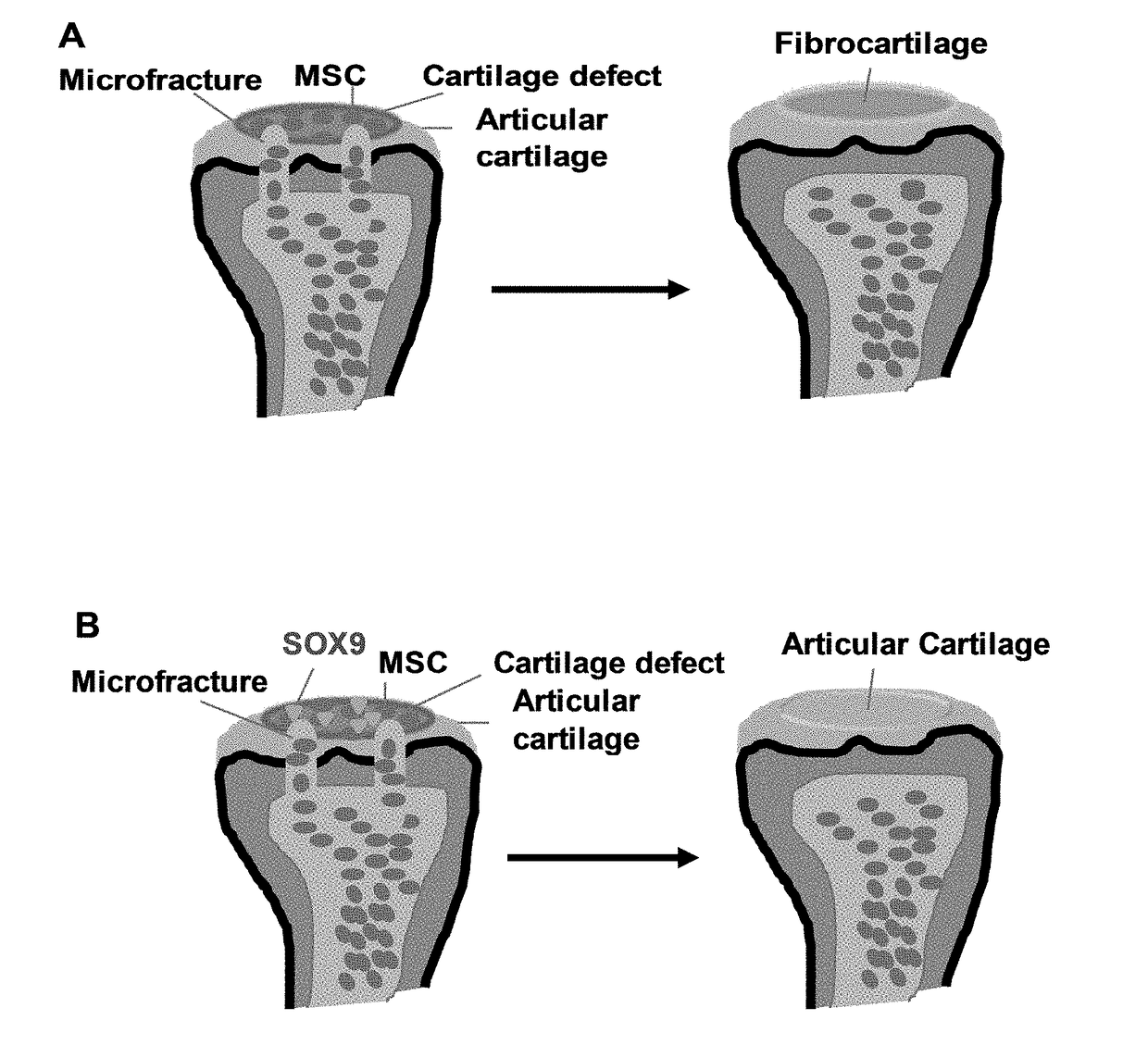
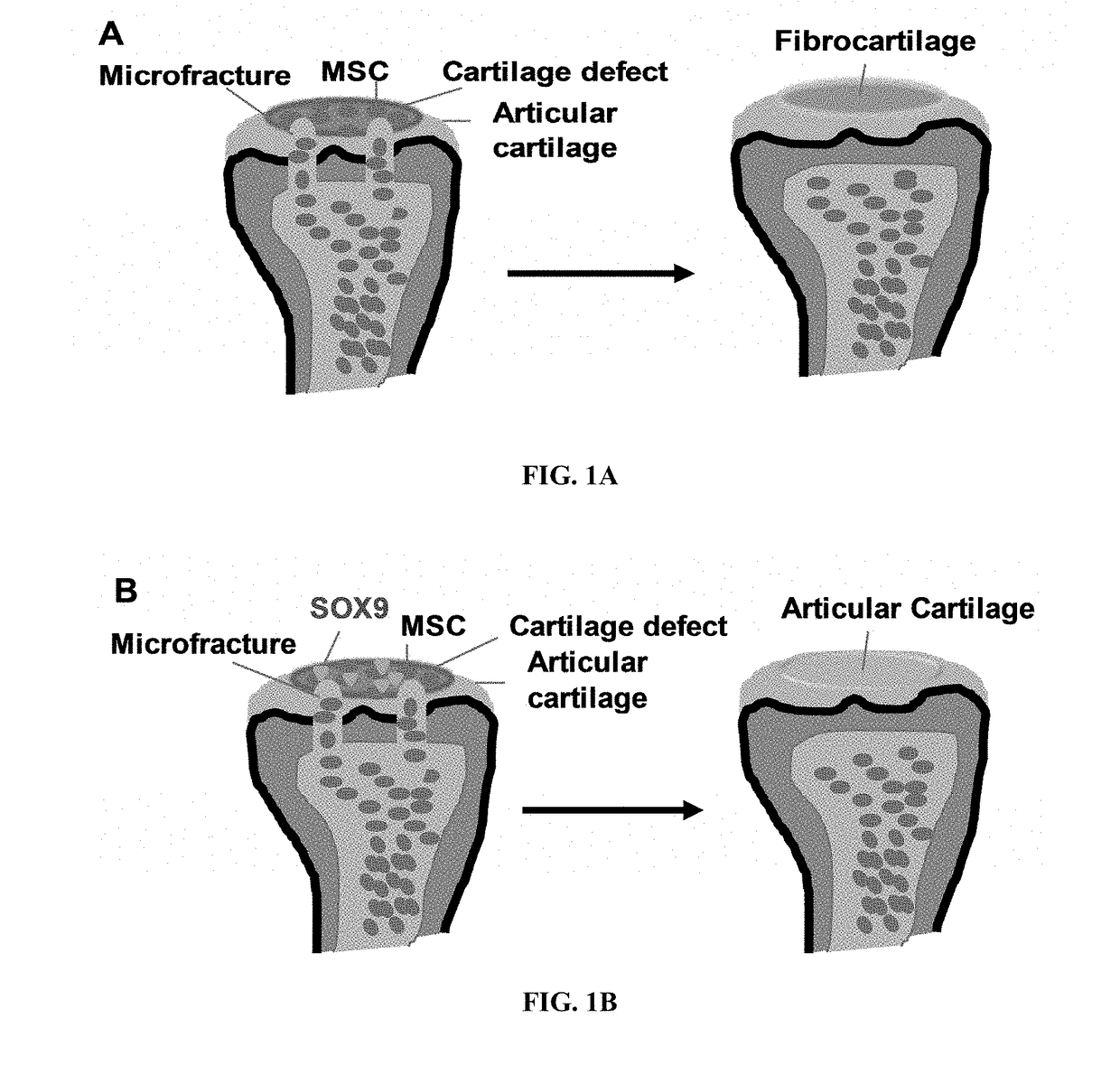
However, like other surgical procedures, the cartilage formed is fibro-cartilaginous.
Fibrocartilage is non-durable and functionally inadequate in the long-term.
Fibrocartilage has poor resistance to shear forces, in contrast to hyaline cartilage.
Hyaline cartilage has a poor intrinsic capacity for healing.
As the presence of type I collagen impairs cartilage-specific matrix architecture and mechanical function, repair of cartilage damage by fibrocartilage leads to morbidity and functional impairment.
Healing of cartilage damage with hyaline cartilage rather than fibrocartilage remains a challenging clinical problem.

Method used


Image

  • Methods for repairing cartilage damage
  • Methods for repairing cartilage damage
  • Methods for repairing cartilage damage

Examples


example 1

[0056]Super-charged SOX9 (scSOX9) comprising SOX9 protein fused with super-charged green fluorescence protein (scGFP) can penetrate MSCs in vitro.

[0057]Method

[0058]Commercial human MSCs (ScienCell Research Laboratories) at passage 5 were maintained and expanded in culture medium in sub-confluence condition. Induction of MSC differentiation was carried out in high-throughput cell aggregate culture as described. 2.5×10⁵ cells / well MSC cells in 0.2 ml are cultured in V-bottomed polypropylene 96-well plates. For positive control, MSCs were cultured in DMEM-HG supplemented with 10% ITS+Premix Tissue Culture Supplement (Becton Dickson), 10−7 M dexamethasone and 10 ng / ml TGF-β1. Under this culture condition, MSCs undergo chondrogenic differentiation within 2-3 weeks, producing abundant extracellular matrix composed primarily of cartilage-specific molecules such as type II collagen and aggrecan. The expression of these cartilage markers were used as evidence of the chondrogenic differentiat...

example 2

[0064]Super-charged SOX9 (scSOX9) comprising SOX9 protein fused with super-charged green fluorescence protein (scGFP) can induce chondrogenesis in vivo.

[0065]The release of scSOX9 from carrier was tested. A commercial bilayer collagen membrane (Bio-Gide) was used to serve as a carrier for scSOX9 to be administered at the site of microfracture. A Bio-Gide membrane at 4 mm in diameter was soaked in 100 μg / ml of scSOX9 solution for one hour. Green fluorescence was grossly visible on the Bio-Gide membrane, and 60% of the total scSOX9 was carried. Release of scSOX9 from Bio-Gide membrane was tested by rinsing the membrane with PBS containing 20 U / ml heparin (pH7.4) for 1 hour, over 95% of scSOX9 bound on Bio-Gide membrane was released and re-dissolved in solution.

[0066]The efficiency of scSOX9 delivery into MSC in vivo was assessed. A cylindrical cartilage defect of 4 mm in diameter and 3 mm in depth was created in patellar groove of the femur of New Zealand female rabbits. Microfracture...

example 3

[0068]Design, construct and screen cell-penetrating, non-immunogenic SOX9 protein variants for reprogramming MSC to chondrocytes in vitro.

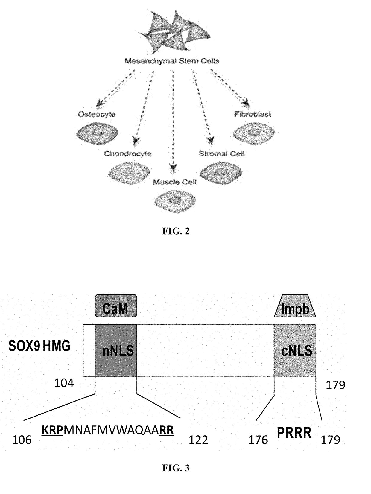
[0069]The ability of scGFP fused proteins to efficiently penetrate into cells depends on the strong positive charge of the scGFP moiety, or the fusion proteins' net theoretical charge to molecular weight ratio. Another way to make the SOX9 protein transducible is to add a cell-penetrating peptide (CPP) to SOX9. Upon studying its sequence using the support vector machine (SVM)-based model, an internal putative CPP (177YQPRRRKSVK186) has been identified embedded in SOX9 (Table 1). Therefore, it is possible that the native SOX9 itself is capable of penetrating into cells without the help of the scGFP moiety. Coincidently, this putative CPP also contains the cNLS as shown in FIG. 3.

[0070]To further enhance the strength of the putative CPP and improve the ability of SOX9 transduction, a series of SOX9 variants are constructed by initially replacing ind...


PUM

PropertyMeasurementUnit
Compositionaaaaaaaaaa
Biocompatibilityaaaaaaaaaa
Bioabsorbableaaaaaaaaaa

Abstract

A method for repairing cartilage damage comprising (a) creating a microfracture or performing other bone marrow stimulation techniques on a patient inflicted with cartilage damage; and (b) administering a composition to the microfracture, wherein the composition comprises an agent capable of regenerating organized hyaline cartilage.

Description

CROSS-REFERENCE TO RELATED APPLICATIONS[0001]This application claims priority to U.S. provisional patent application No. 62 / 008,513, filed Jun. 6, 2014, the disclosure of which is incorporated herein by reference in its entirety.FIELD OF THE INVENTION[0002]The present invention generally relates to methods for repairing cartilage damage.BACKGROUND OF THE INVENTION[0003]Articular cartilage is a highly organized tissue with low cell density and limited nutrient supply. Once it is damaged by trauma or degenerative arthritis, it has a limited capacity for regeneration. The most common joint disorder, osteoarthritis (OA), is afflicting millions of people with symptoms including severe pain, swelling and clicking of joints. To make things worse, OA cannot be cured—only its symptoms can be controlled.[0004]OA is the most common form of arthritis and the fourth leading cause of disability worldwide. Over 70% of Americans between the ages of 55 and 70 are estimated to be affected by OA. Trea...

Claims


Application Information

Patent Timeline
IPC IPC(8): A61L27/38A61K9/00A61L27/54A61L27/24A61K38/17C07K14/47
CPCA61L27/3852A61K38/1709C07K14/47A61L27/54A61L27/24A61L2300/412C07K2319/095C07K2319/60C07K2319/10A61L2430/06A61K9/0024C07K2319/00A61K9/06A61K47/42A61L27/225A61L27/3834A61L27/58A61L2300/25A61L27/227A61P19/00A61P37/06A61P43/00
Inventor CHU, CONG-QIUZHU, YONGWU, SHILIBAO, JUN
Owner VIVOSCRIPT
Learn More