
Device and method for purifying virally infected blood

a technology of viral infection and purification method, which is applied in the field of therapeutic methodologies and devices for treating viral infections, can solve the problems of limiting the effectiveness of therapy, increasing the likelihood of viral infection with these deadly agents, and difficulty in treatment for viral diseases, so as to reduce the antigenic assault on the immune system

Inactive Publication Date: 2016-01-07
AETHLON MEDICAL INC
View PDF1 Cites 4 Cited by
  • Summary
  • Abstract
  • Description
  • Claims
  • Application Information

AI Technical Summary

Benefits of technology

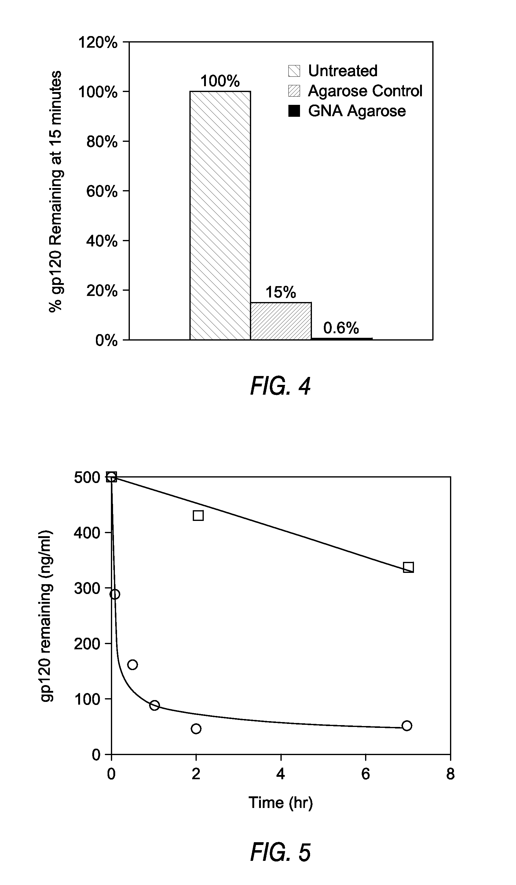
[0016]The present invention utilizes lectins to bind, immobilize and retain whole virus, particularly infectious virus, as well as parts thereof, thus allowing a diminution of circulating virus and potential reduction of antigenic assault on the immune system. Of particular interest is the ability to preferentially remove live or infectious viral particles as compared to total viral load as measured, for example, by PCR.

Problems solved by technology

Aside from natural infection, the emerging threat of bioterror makes mass infections with these deadly agents ever more likely.
Therapy is difficult for viral diseases as antibiotics have no effect on viruses and few antiviral drugs are known.
In cases where drug treatments are available, the occurrence of resistant mutations and drug side effects often limit the effectiveness of therapy.
The best way to prevent viral diseases is through vaccination; however, vaccines are unavailable for a large number of viruses, including many of the viruses listed above.
Although there are vaccines present for others, many available vaccine strategies are either not fully effective, as in the case of Hepatitis B Virus, or present potentially life-threatening side-effects, such as the vaccine released and recalled for rotavirus.
Further, where vaccines do exist they are predominantly preventive and largely ineffective once a viral infection becomes established in the host.
A small proportion of DHF cases lead to dengue shock syndrome (DSS) which has a high mortality rate.
There are no vaccines available to stem outbreak and the only therapy available is a sit and wait approach.
The hallmark of AIDS is the loss of CD4+ T cells, which ultimately leaves the immune system unable to defend against opportunistic infections.
They do not directly remove HIV virus.
However, none of these treatments effectively remove both virus and viral proteins.
However, none of these chromatographic materials are selective for viruses and will clearly remove many other essential substances.
Thus they are not useful for in vivo blood purification.
However, this strategy was inefficient as it required extracorporeal absorption of blood and did not provide for a mechanism to remove free HIV viral particles from the blood (Lopukhin et al., 1991, supra).
Accordingly, although lectins are known to bind viral envelope glycoproteins, no previous technologies have demonstrated the ability to directly adsorb a wide spectrum of viruses, preferably enveloped viruses, from the blood using lectins in the setting of ex vivo dialysis or plasmapheresis.

Method used


Image

  • Device and method for purifying virally infected blood
  • Device and method for purifying virally infected blood
  • Device and method for purifying virally infected blood

Examples


example 1

[0082]This Example demonstrates the preparation of an affinity matrix using GNA covalently coupled to agarose using cyanogen bromide. Cyanogen bromide (CNBr) activated agarose was used for direct coupling essentially according to Cuatrecasas, et al (Cuatracasas et al. Proc Natl Acad Sci USA 61(2): 636-643, 1968). In brief, 1 ml of GNA at a concentration of 10 mg / ml in 0.1M NaHCO3 pH 9.5 was added to 1 ml CNBr activated agarose (Sigma, St. Louis, Mo.) and allowed to react overnight in the cold. When the reaction was complete, unreacted materials were aspirated and the lectin coupled agarose washed extensively with sterile cold PBS. The lectin agarose affinity matrix was then stored cold until ready for use. Alternatively, GNA agarose is available commercially from Vector Labs (Burlingame, Calif.)

example 2

[0083]This Example demonstrates preparation of the lectin affinity matrix using GNA covalently coupled to glass beads via Schiff's base and reduction with cyanoborohydride. The silica lectin affinity matrix was prepared by a modification of the method of Hermanson (Hermanson. Bioconjugate Techniques: 785, 1996). GNA lectin was dissolved to a final protein concentration of 10 mg / ml in 0.1M sodium borate pH 9.5 and added to aldehyde derivatized silica glass beads (BioConnexant, Austin Tex.). The reaction is most efficient at alkaline pH but will go at pH 7-9 and is normally done at a 2-4 fold excess of GNA over coupling sites. To this mixture was added 10 μl 5M NaCNBH3 in 1N NaOH (Aldrich, St Louis, Mo.) per ml of coupling reaction and the mixture allowed to react for 2 hours at room temperature. At the end of the reaction, remaining unreacted aldehyde on the glass surfaces are capped with 20 μl 3M ethanolamine pH 9.5 per ml of reaction. After 15 minutes at room temperature, the react...

example 3

[0084]This Example demonstrates preparation of GNA covalently coupled to aminocelite using glutaraldehyde. Aminocelite was prepared by reaction of celite (silicate containing diatomaceous earth) by overnight reaction in a 5% aqueous solution of aminopropyl triethoxysilane. The aminated celite was washed free of excess reagent with water and ethanol and dried overnight to yield an off white powder. One gram of the powder was then suspended in 5 ml 5% glutaraldehyde (Sigma) for 30 minutes. Excess glutaraldehyde was then removed by filtration and washing with water until no detectable aldehyde remained in the wash using Schiffs reagent. The filter cake was then resuspended in 5 ml of Sigma borohydride coupling buffer containing 2-3 mg / ml GNA and the reaction allowed to proceed overnight at room temperature. At the end of the reaction, unreacted GNA was washed off and the unreacted aldehyde aminated with ethanolamine as described. After final washing in sterile PBS, the material was sto...


PUM


Abstract

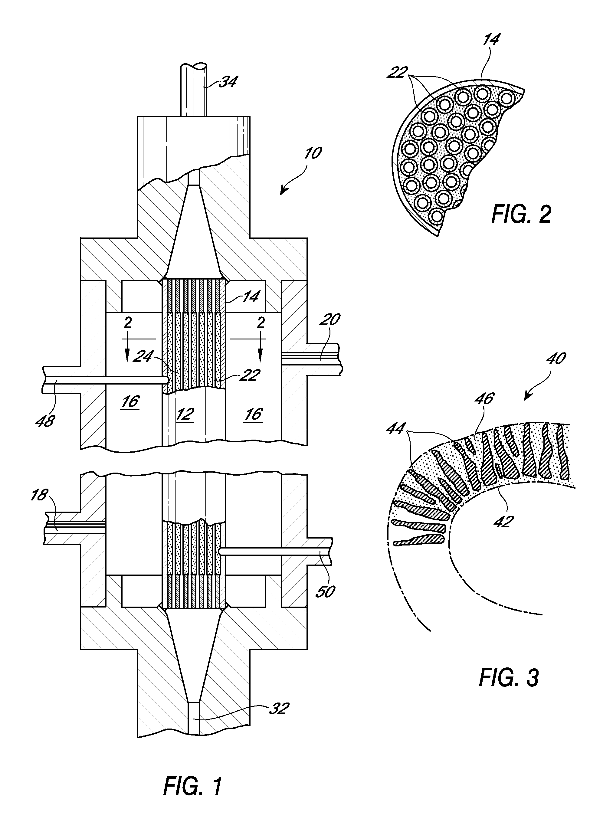
The present invention relates to a method for using lectins that bind to pathogens having surface glycoproteins or fragments thereof which contain glycoproteins, to remove them from infected blood or plasma or other fluids in an extracorporeal setting. Accordingly, the present invention provides a methods and devices for reducing viral load or plaque forming units in blood or plasma from one or more infected individuals. A preferred embodiment of the method comprises passing the blood or plasma through a porous hollow fiber membrane wherein lectin molecules are disposed proximate to the membrane, collecting pass-through blood or plasma and optionally reinfusing the pass-through blood or plasma into the individual. Additionally, the present invention provides a methods and devices for the reduction of plaque forming units, cleared more rapidly and more efficiently than overall viral load.

Description

RELATED APPLICATIONS[0001]This application is a continuation of U.S. application Ser. No. 12 / 600,236, filed May 12, 2011, which is the U.S. National Phase under 35 U.S.C. §371 of International Application No. PCT / US2008 / 063946, filed May 16, 2008 under the Patent Cooperation Treaty (PCT), which was published by the International Bureau in English, which designates the United States and claims the benefit of U.S. Provisional Application No. 60 / 938,432, filed May 16, 2007, the disclosures of which are hereby expressly incorporated by reference in their entireties.BACKGROUND OF THE INVENTION[0002]1. Field of the Invention[0003]The present invention relates to the field of therapeutic methodologies and devices for treating viral infections and removing viral particles from contaminated fluids.[0004]2. Description of the Related Art[0005]A large number of viruses have been described which are pathogenic for humans. Viruses such as ebola, marburg, smallpox, lassa, dengue, influenza (e.g. ...

Claims


Application Information

Patent Timeline
Patent Type & Authority Applications(United States)
IPC IPC(8): A61M1/36B01D15/22B01D15/38B01D61/02B01D69/02B01D69/08
CPCA61M1/362B01D61/027B01D15/3823B01D2325/02B01D69/08B01D69/02A61M2202/206B01D15/22A61M1/3679A61M2202/097A61M2205/273A61M1/3486A61P31/14A61P31/16A61P31/18A61P31/20
Inventor TULLIS, RICHARD H.HANDLEY, HAROLD H.JOYCE, JAMES A.DUFFIN, PAUL
Owner AETHLON MEDICAL INC
Learn More