
Micropatterned co-culture systems as infectious disease analysis platforms

a co-culture system and infectious disease technology, applied in the field of micropatterned co-culture systems as infectious disease analysis platforms, can solve the problems of difficult realization of goals, severe side effects of ribavirin, and inability to cure many infected patients, and achieve the effect of high throughput experimentation and automated optical readou

Inactive Publication Date: 2015-09-24
MASSACHUSETTS INST OF TECH
View PDF1 Cites 3 Cited by
  • Summary
  • Abstract
  • Description
  • Claims
  • Application Information

AI Technical Summary

Benefits of technology

This patent describes a new method for creating and using cell cultures that can be used for drug discovery and vaccine development. These cell cultures can be infected with different viruses, like HCV, to study their spread and infection. The method also allows for the creation of reporter systems that can help evaluate the effectiveness of different treatments. The cultures can be used with patient isolates from actual infections, making them useful for studying in vivo infections. The method can also be applied to different models of pathogenesis, making it applicable to a variety of tissue types and animal models. Overall, this invention provides a new tool for researchers to better understand the mechanisms of viral infections and develop new treatments.

Problems solved by technology

Current drug therapies (pegylated interferon and ribavirin) have severe side effects and do not cure many infected patients even after lengthy (months to year) treatment regimens.
However, these goals have been difficult to realize partly due to the difficulties in maintaining sustained and reproducible infection of isolated primary human hepatocytes (main cell type of the liver) with HCV in vitro.
However, this cancer-derived cell line has abnormal repertoire and levels of liver-specific functions.
Monitoring molecular biology of the parasite in the liver is difficult using current methods, and there is no easy way to determine in cell culture whether the vaccine is properly arresting the parasite.
Historically cell culture techniques and tissue development failed to take into account the necessary microenvironment for cell-cell and cell-matrix communication as well as an adequate diffusional environment for delivery of nutrients and removal of waste products.
While many methods and bioreactors have been developed to grow tissue for the purposes of generating artificial tissues for transplantation or for toxicology studies, these bioreactors do not adequately simulate, in vitro, the mechanisms by which nutrients, gases, and cell-cell interactions are delivered and performed in vivo.
Thus, cell culture systems and bioreactors that do not simulate these in vivo delivery mechanisms do not provide a sufficient corollary to in vivo environments to develop tissues or measure tissue responses in vitro.
Further, traditional cell culture systems often fail to provide adequate information on the liver toxicity and bioavailability of drug candidates.
These issues have caused 50% of new drug candidates to fail in Phase I clinical trials.
Also, a third of drug withdrawals from the market and more than half of all warning labels on approved drugs are primarily due to adverse affects on the liver.
Current in vitro liver models used by the pharmaceutical industry, though useful in a limited capacity, are not fully predictive of in vivo liver metabolism and toxicity.
Thus, research has increasingly turned towards using isolated primary human hepatocytes as the gold standard for in vitro studies; however, hepatocytes are notoriously difficult to maintain in culture as they rapidly lose viability and phenotypic functions.
Nevertheless, monitoring of HCV infection and treatment poses specific challenges in cell culture techniques.
Firstly, HCV has a low infectivity of cells in culture, making the preparation of in vitro culture systems difficult or impossible.
Secondly, monitoring HCV replication is destructive, as the cells are typically destroyed before assessing HCV RNA levels.

Method used


Image

  • Micropatterned co-culture systems as infectious disease analysis platforms
  • Micropatterned co-culture systems as infectious disease analysis platforms
  • Micropatterned co-culture systems as infectious disease analysis platforms

Examples


example 1

Disease Platform for HCV Drug Development

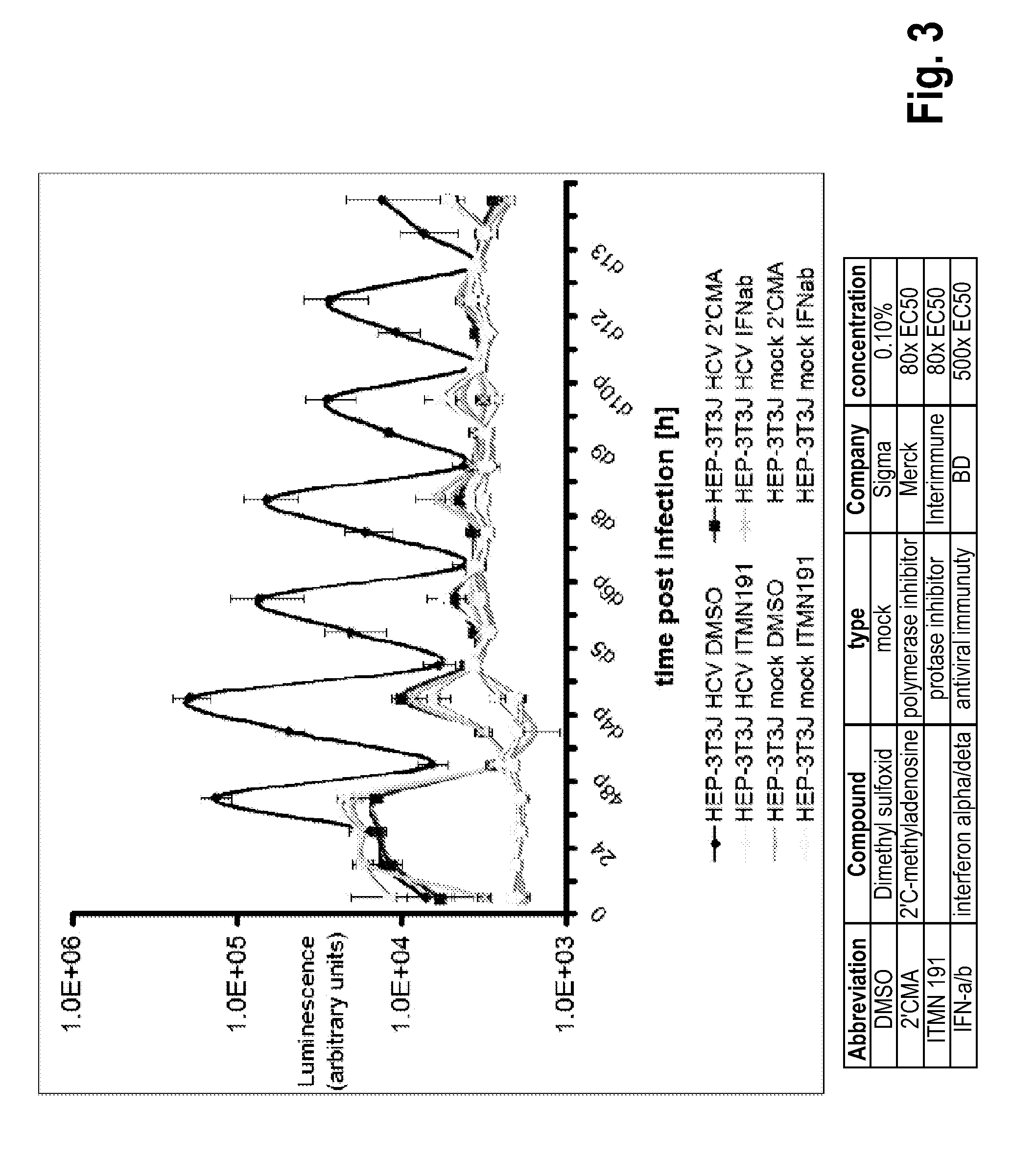
[0178]This Example presents the development and optimization of a disease platform for HCV drug development that couples micropatterned co-cultures of primary human hepatocytes and stromal cells in a multi-well format (FIG. 1) with highly sensitive viral reporter systems and optimized treatment protocols. The developed human liver platform can: a) Support entry of HCV-like particles via receptor-mediated processes (FIG. 2), and b) Produce and release infectious HCV particles into the culture supernatant for several weeks in vitro, and that such release can be blocked by treatment of cultures with small molecule inhibitors of viral proteins (FIG. 3). It was also found that infection of micropatterned co-cultures is dependent on the dose and time of exposure to the initial inoculum (FIG. 4), and magnitude of HCV infection in micropatterned co-cultures is 4-6 fold higher than in pure hepatocyte monolayers, the current gold standard in the field ...

example 2

A Fluorescent-Based Reporter for HCV Infection

[0180]A novel cell-based fluorescent reporter system has been developed to detect infection of unmodified HCV genomes. The example system described here uses an HCV-dependent fluorescence relocalization (HDFR) cassette that comprises a fluorescent protein (e.g. EGFP, mCherry or TagRFP), an SV40 nuclear localization sequence (NLS), and a C-terminal mitochondrial-targeting domain (IPS) derived from the interferon-beta promoter stimulator 1 protein, IPS-1. IPS-1 is a known cellular substrate for the HCV NS3-4A protease, and IPS-1 mutation C508Y has been shown to abolish cleavage. In HCV-infected cells the HDFR cassette is processed by the viral NS3-4A protease, resulting in translocation of the fluorescent protein from the mitochondria to the nucleus. Using a lentivirus based expression system to stably express the HDFR cassette, we have successfully established this system in the highly HCV permissive human hepatoma cell line, Huh-7.5 and ...

example 3

Live Cell Imaging of HCV Infection

[0185]Live cell imaging of HCV infection was demonstrated using the Huh7.5 cell line stably expressing a modified red fluorescent version of EGFP-IPS (Tag-RFP-nls-IPS) and mito-EGFP. TagRFP-nls-IPS encodes TagRFP (a bright red fluorescent protein), a SV40 nuclear localization sequence (nls), and the mitochondrial targeting sequence from the C-terminus of IPS-1. Mito-EGFP encodes the mitochondrial targeting sequence from subunit VIII of cytochrome-c oxidase. Localization of Mito-EGFP to the mitochondria, shown as green staining surrounding cell nuclei. Following HCVcc infection of the resultant cell line, relocalization of the TagRFP-nls-IPS from the mitochondria to the nucleus was visualized (red / violet staining of cell nuclei) as a result of cleavage by the HCV NS3 / 4A protease and subsequent transport to the nucleus via the nls. Mito-EGFP localization is unaltered by HCV infection and serves as a marker for the mitochondria. The stark visualization...


PUM

PropertyMeasurementUnit
concentrationsaaaaaaaaaa
distanceaaaaaaaaaa

Abstract

Cell cultures are provided that include a population of micropatterened hepatocytes and one or more non-parenchymal cell populations, where the hepatocytes are infected with a virus or parasite and include a reporter of virus or parasite infection. Methods of making and using the cell cultures are also provided.

Description

RELATED APPLICATIONS[0001]This application is a divisional of U.S. Utility application Ser. No. 13 / 003,214, filed Jul. 5, 2011, which claims priority to International Application No. PCT / US2009 / 049849, filed Jul. 7, 2009, which claims priority to U.S. provisional application Ser. No. 61 / 078,683 filed Jul. 7, 2008 and U.S. provisional application Ser. No. 61 / 174,449 filed Apr. 30, 2009. The entire contents of each of the foregoing applications are incorporated herein by this reference.BACKGROUND[0002]More than 150 million people worldwide are infected with the Hepatitis C virus (HCV), and about 75% of those infected display chronic infection, which can lead to liver cirrhosis and hepatocarcinoma. Current drug therapies (pegylated interferon and ribavirin) have severe side effects and do not cure many infected patients even after lengthy (months to year) treatment regimens. Hence, there is an urgent need to better understand the variability in host response and propensity for chronic ...

Claims


Application Information

Patent Timeline
Patent Type & Authority Applications(United States)
IPC IPC(8): G01N33/50C12Q1/18
CPCG01N33/5067C12Q1/18G01N33/5014C12N2535/10
Inventor BHATIA, SANGEETA N.KHETANI, SALMAN R.
Owner MASSACHUSETTS INST OF TECH
Learn More