
Magnetic graphene-like nanoparticles or graphitic nano- or microparticles and method of production and uses thereof

a technology of graphene-like nanoparticles and graphene-like nanoparticles, which is applied in the field of magnetic graphene-like nanoparticles or graphitic nanoor microparticles and their production methods, and can solve the problems of unexplored potential and efficacy of mnsup>2+/sup>-ion carbon nanostructure complexes as mri cas, and the ion system of carbon nanostructure-gd

Active Publication Date: 2014-06-12
THE RES FOUND OF STATE UNIV OF NEW YORK
View PDF2 Cites 22 Cited by
  • Summary
  • Abstract
  • Description
  • Claims
  • Application Information

AI Technical Summary

Benefits of technology

The present invention provides a magnetic composition that includes a magnetic metal and a graphene-like carbon nanostructure, such as a carbon nanoplatelet or carbon nanoribbon, which exhibits a high relaxivity and can be used for magnetic resonance imaging (MRI) of a subject. The magnetic composition can also be water solubilized and attached to a graphene-like nanostructure. The carbon nanoplatelet or carbon nanoribbon can have a thickness of about 20 nm or less. The magnetic metal can be a room temperature paramagnetic metallic element, a room temperature ferromagnetic metallic element, or a rare earth metal. The magnetic metal can be intercalated in the graphene-like nanostructure or metal compound. The magnetic composition can be used in MRI imaging and can be produced using a method involving oxidizing graphite with a mixture of sulfuric acid, nitric acid, manganese chloride, and potassium permanganate, and sonicating a suspension of the product obtained. The magnetic composition can also be water solubilized using a method known in the art.

Problems solved by technology

However, the potential and efficacy of Mn2+-ion carbon nanostructure complexes as MRI CAs still has not been investigated.
A major reason for this lack of understanding is that unlike Gd3+ ion chelates, which can be prepared at a very high level of purity and unambiguously characterized, the carbon nanostructure-Gd3+ ion systems are rather complex mainly due to their particulate nature, and intricate relationships linking their chemical, geometric, and magnetic characteristics to their properties as MRI contrast agents.
Additionally, to date, there have been no studies performed to systematically investigate whether the high increase in relaxivity and unconventional NMRD profiles are unique to paramagnetic ions confined in single-walled carbon nanotubes, which are seamless cylinders formed from a graphene sheet, or in general observed for paramagnetic ions confined in other graphene or graphitic structures.

Method used


Image

  • Magnetic graphene-like nanoparticles or graphitic nano- or microparticles and method of production and uses thereof
  • Magnetic graphene-like nanoparticles or graphitic nano- or microparticles and method of production and uses thereof
  • Magnetic graphene-like nanoparticles or graphitic nano- or microparticles and method of production and uses thereof

Examples


example 1

Materials and Methods

[0082]Graphene Nanoplatelet (GNP) Synthesis:

[0083]Oxidized graphite was prepared from analytical grade micro-graphite (45 μm, 496596-Sigma Aldrich) by modified Hummer's method (Geng, Y., Wang, S. & Kim, J. Preparation of graphite nanoplatelets and graphene sheets. Journal of colloid and interface science 336, 592-598 (2009); Hummers Jr, W. & Offeman, R. Preparation of graphitic oxide. Journal of the American Chemical Society 80, 1339-1339 (1958)). In a typical exfoliation procedure, dried oxidized graphite (200 mg) was suspended in a round bottom flask containing water (200 ml), along with MnCl2 and sonicated for 1 h in an ultrasonic bath cleaner (Fischer Scientific, FS60, 230W) (Stankovich, S. et al. Synthesis of graphene-based nanosheets via chemical reduction of exfoliated graphite oxide. Carbon 45, 1558-1565 (2007)). 50 ml of this uniform solution was centrifuged and pellet was dried overnight to obtain oxidized graphene nanoplatelets (GNPs). The remaining 1...

example 2

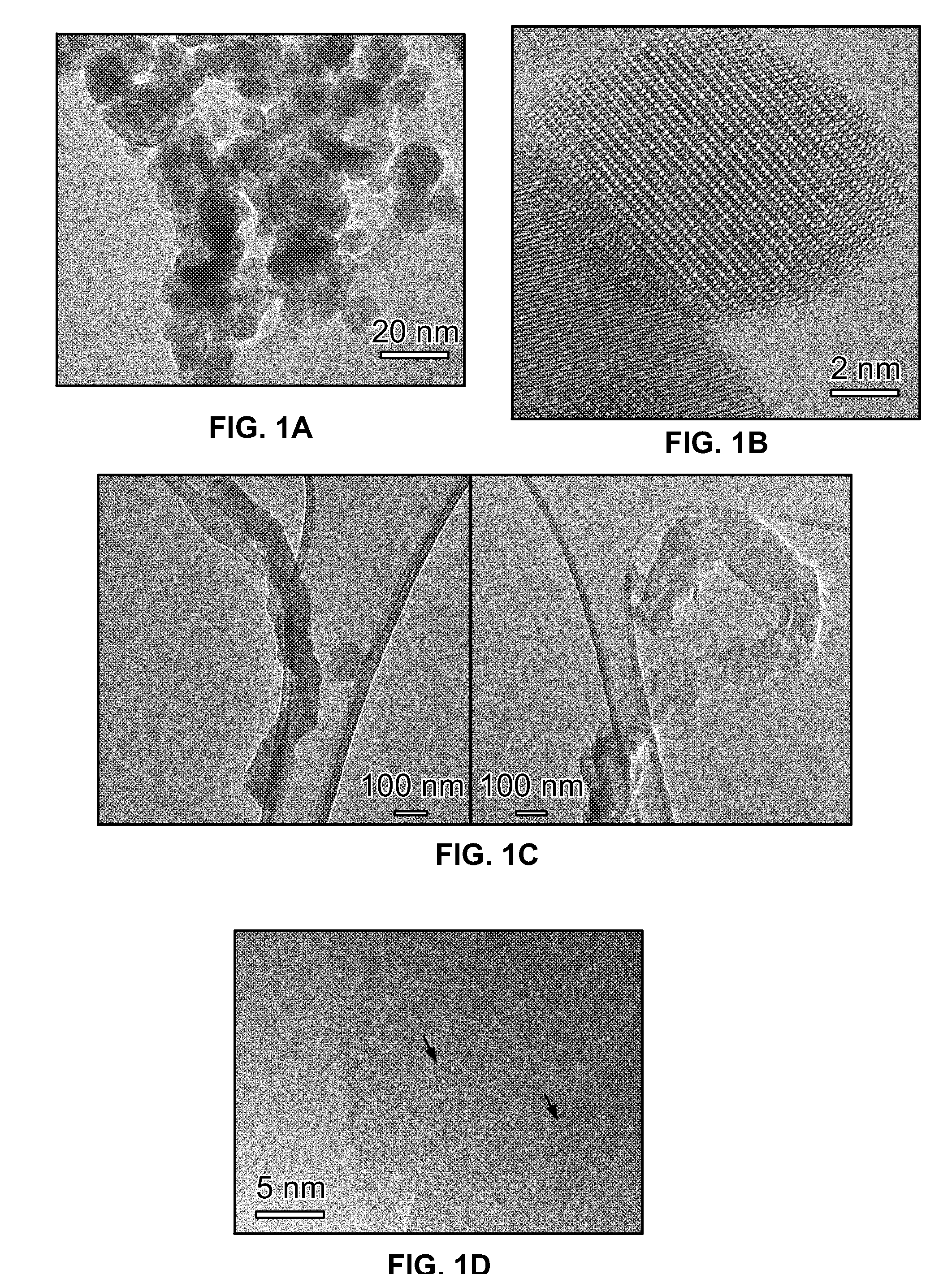
[0111]The novel graphene nanoplatelets (GNPs; small stacks (1-12 layers) of graphene (one-atom-thick sheets of graphite) with thickness between 1 to 5 nanometers (nm) and diameters of ˜50 nm) (FIGS. 1a and 1b) was tested as MRI contrast agent in MRI scans of a mouse. The GNPs were monodispersed, water-soluble, intercalated (insertion of chemical species within the voids between two graphene sheets), and coordinate with trace amounts of manganese (0.1% w / w (w=weight) of Manganese in GNPs, i.e. 0.1 gram of manganese per 100 gram of GNPs). The GNPs showed relaxivity of r1=45 mM−1s−1 which is nearly 16 times higher than Mn-DPDP (Teslascan, clinical Mn-based CA, r1=2.8 mM−1s−1 at 3 T) and ˜10 times greater than Gd-DTPA (Magnevist, Clinical Gd-based CA, r1=4.2 mM−1s−1 at 3 T). Scans using T1 weighted small animal MRI using a 3 Tesla clinical scanner (FIG. 6) showed ˜100 times greater contrast enhancement compared to Magnevist at clinical dosages.

example 3

Graphene Nanoplatelets (GNPs)

[0112]First, graphite oxide (GO) is prepared from analytical grade micro-graphite using modified Hummer's method (treatment with potassium permanganate and concentrated surphuric acid) and converted to graphene nanoplatelets (GNPs) by sonication for 1 h in an ultrasonic bath cleaner (Stankovich, S.; Dikin, D. A. et al. Nature 2006, 442, 282). Next, the GNPs is water solubilized using a synthesis protocol that is similar to a cycloaddition reaction used to add carboxylic acid functionalities across carbon-carbon double bonds of fullerenes and metallofullerenes (Sithamaran, B.; Zakharian, T. Y. et al. Molecular Pharmaceutics 2008, 5, 567).). The GNPs is water solubilized by covalently or non-covalently coating them with natural polymers such as dextran and synthetic polymers such as polyethylene glycol.

[0113]Controls

[0114]Magnevist is used in experiments where controls are needed, since it is considered the benchmark for clinical MRI contrast media. Additi...


PUM

PropertyMeasurementUnit
thicknessaaaaaaaaaa
thicknessaaaaaaaaaa
thicknessaaaaaaaaaa

Abstract

A magnetic graphene-like nanoparticle or graphitic nano- or microparticle exhibits a high relaxivity, and is useful as a MRI contrast agent. A composition for use with MRI imaging, comprising a sufficient amount of the magnetic graphene-like nanoparticles or graphitic nano- or microparticles and one or more physiologically acceptable carriers or excipients. Methods of using the magnetic graphene-like nanoparticles or graphitic nano- or microparticles as MRI contrast agents. Methods of producing the magnetic graphene-like nanoparticle or graphitic nano- or microparticle.

Description

REFERENCE TO RELATED APPLICATIONS[0001]This application claims priority to U.S. Provisional Application No. 61 / 483,309, filed May 6, 2011, the content of which is incorporated herein by reference in its entirety.FIELD OF THE INVENTION[0002]The present invention relates to magnetic graphene-like nanoparticles or graphitic nano- or microparticles and method of production thereof. The present invention also relates to methods of using the magnetic graphene-like nanoparticles or graphitic nano- or microparticles as MRI contrast agents.BACKGROUND OF THE INVENTION[0003]MRI is primarily used to non-invasively render anatomical details for improved diagnosis of many pathologies and diseases (Sitharaman, B. & Wilson, L. J. Gadofullerenes and Gadonanotubes: A new paradigm for high-performance magnetic resonance imaging contrast agent probes Journal of Biomedical Nanotechnology 3, 342-352 (2007); Pan, D. et al. Revisiting an old friend: manganese-based MRI contrast agents. WIREs Nanomedicine a...

Claims


Application Information

Patent Timeline
Patent Type & Authority Applications(United States)
IPC IPC(8): A61K49/10
CPCA61K49/10C01B2204/20B82Y30/00B82Y40/00B82Y15/00C01B32/184C01B32/192A61K49/0095C01P2004/24C01P2004/17C01P2006/42
Inventor SITHARAMAN, BALAJIPARATALA, BHAVNA S.
Owner THE RES FOUND OF STATE UNIV OF NEW YORK
Learn More