Method for in vitro detecting keratin gene fusion of squamous-cell cancer
a keratin gene and in vitro technology, applied in the field of cancer detection methods, can solve problems such as false negative errors, and achieve the effect of high accuracy
- Summary
- Abstract
- Description
- Claims
- Application Information
AI Technical Summary
Benefits of technology
Problems solved by technology
Method used
Image

Examples
embodiment i
Sequencing and Popularization Rate of Gene Fusion in Oral Squamous-Cell Cancer
[0049]A. Test Material and Test Method
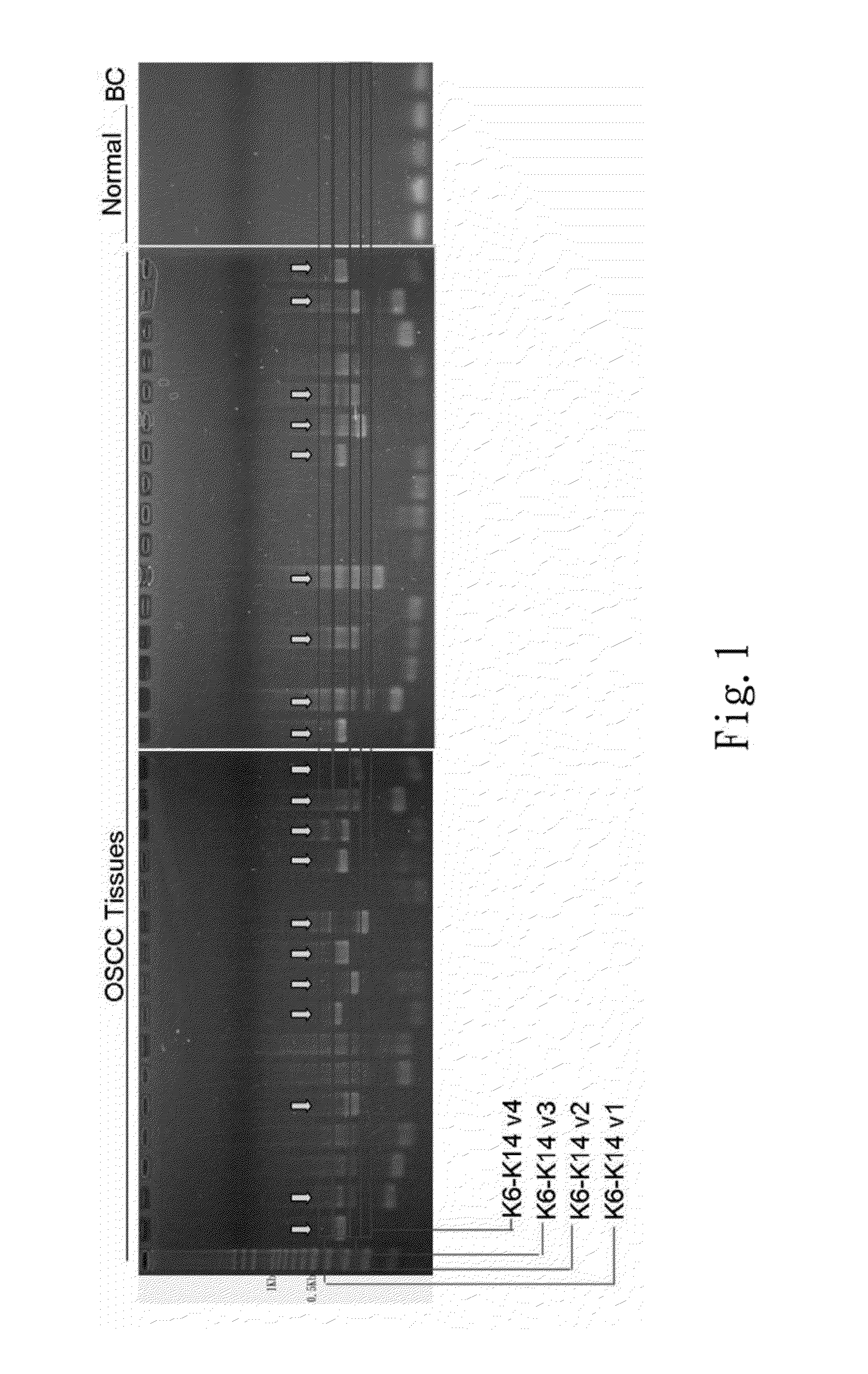
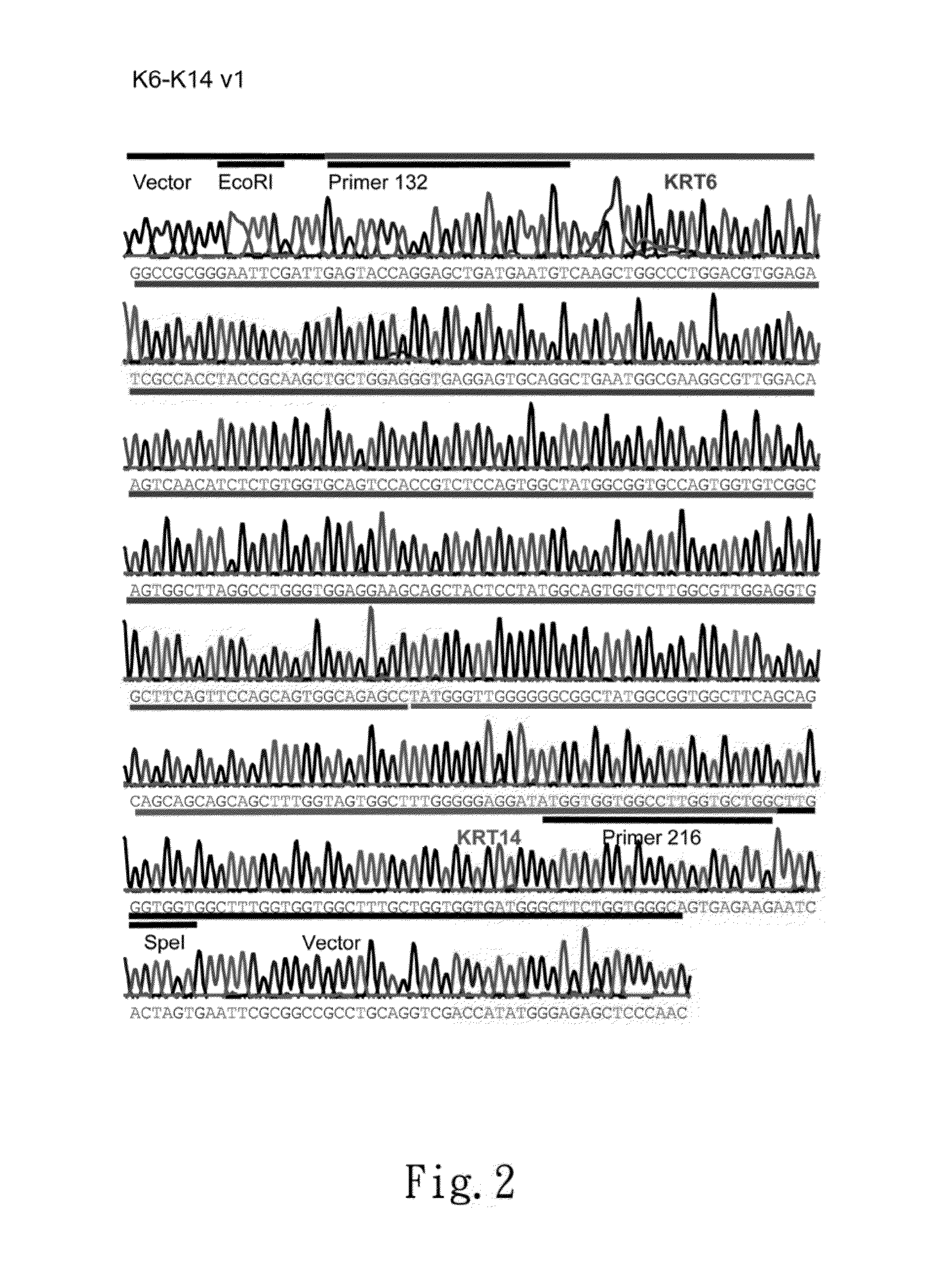
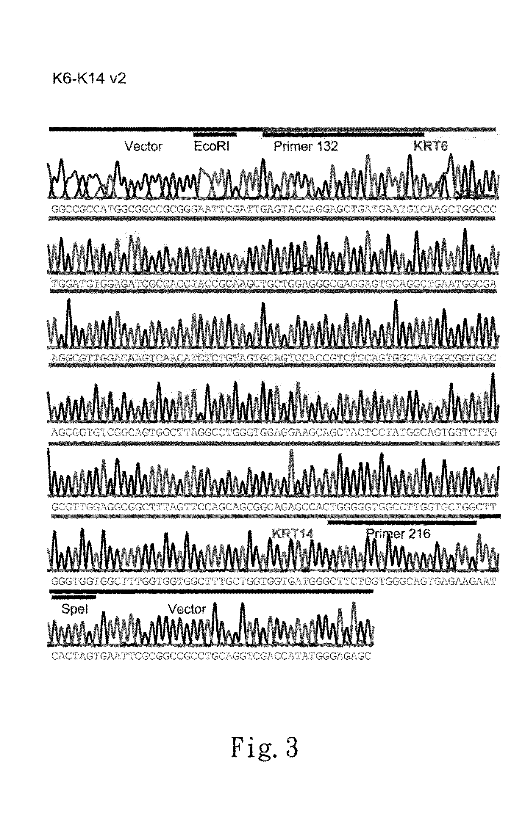
[0050]The test material includes samples of oral squamous-cell cancer (n=48) and normal samples (n=4). All the samples of oral squamous-cell cancer are provided by the tissue bank of the China Medical University Hospital. The RNA of the samples is extracted with the RNeasy mini kit (Qiagen), quantified with the Nanodrop fluorescent absorption method, and analyzed with a gel-electrophoresis method. The high capacity cDNA RT kit (Applied Bioscience) is used to reverse-transcript 1 μg of RNA of each sample into cDNA. The cDNA is diluted by a 0.1×TE buffer solution to have a concentration of 50-80 ng / nl. Use 1 μL of cDNA as the template to undertake PCR with APP (Amyloid beta Precursor Protein) gene sequence (SEQ ID No: 1; SEQ ID No: 2) being the primer. Examine the products of PCR with gel-electrophoresis, and discard the APP-negative samples. Use 1 μL of cDNA taken from ...
embodiment ii
[0053]FISH of the Gene Fusion of the SAT Cell Line of OSCC
[0054]A. Test Material and Test Method
[0055]a. Preparation of Sample Glasses
[0056]The test material includes the SAT cell line of OSCC. Cultivate the SAT cell line of OSCC in a T75 culture box until the cells have occupied 80% of the volume. Add 0.2 ml of EtBr (1 mg / ml) to the cells, and place them still at a temperature of 37° C. for 90 minutes. Add 0.1 ml of colcemid (Gibco) to the cells, and place them still at a temperature of 37° C. for 25 minutes. Collect and centrifugally process the cells, and remove the supernatant. Add 10 ml of 0.56% KCl to the cells, and flush them with water for 15 minutes. Centrifugally process the liquid containing cells, and remove the supernatant. Flush the cells with a solution containing methanol and glacial acetic acid by 3:1 at a temperature of 0° C. three times, and fix them. Spray the fixed cells on clean silane coating slides (Muto pure chemicals). Process the cells with 100% alcohol fo...
embodiment iii
Popularization Rate of Gene Fusion in Cervical Squamous-Cell Cancer
[0063]A. Test Material and Test Method
[0064]The test material includes samples of cervical squamous-cell cancer (n=30), which are provided by the tissue bank of the China Medical University Hospital. The RNA of the samples is extracted with the RNeasy mini kit (Qiagen), quantified with the Nanodrop fluorescent absorption method, and analyzed with a gel-electrophoresis method. The high capacity cDNA RT kit (Applied Bioscience) is used to reverse-transcript 1 μg of RNA of each sample into cDNA. The cDNA is diluted by a 0.1×TE buffer solution to have a concentration of 50-80 ng / nl. Use 1 μL of cDNA as the template to undertake PCR with GAPDH gene sequence (SEQ ID No: 3; SEQ ID No: 4) being the primer. Examine the products of PCR with gel-electrophoresis, and discard the GAPDH-negative samples. Use 1 μL of cDNA taken from each APP-positive sample as the template to undertake PCR with the gene fusion sequence KRT6: KRT14 ...
PUM
| Property | Measurement | Unit |
|---|---|---|
| Acidity | aaaaa | aaaaa |
Abstract
Description
Claims
Application Information
 - R&D
- Intellectual Property
- Life Sciences
- Materials
- Tech Scout
- Unparalleled Data Quality
- Higher Quality Content
- 60% Fewer Hallucinations
Browse by: Latest US Patents, China's latest patents, Technical Efficacy Thesaurus, Application Domain, Technology Topic, Popular Technical Reports.
© 2025 PatSnap. All rights reserved.Legal|Privacy policy|Modern Slavery Act Transparency Statement|Sitemap|About US| Contact US: help@patsnap.com