Eureka AIR delivers breakthrough ideas for toughest innovation challenges, trusted by R&D personnel around the world.

Biodegradable Intravitreal Tyrosine Kinase Implants

a biodegradable, intravitreal technology, applied in the field of eye treatment, can solve the problems of severe restriction of systemic drug penetration into the retina, physical damage to the lens, retinal detachment, etc., and achieve the effect of few or no negative side effects

Active Publication Date: 2014-01-30
ALLERGAN INC
View PDF2 Cites 82 Cited by
  • Summary
  • Abstract
  • Description
  • Claims
  • Application Information

AI Technical Summary

Benefits of technology

The present invention provides new drug delivery systems for extended or sustained drug release in the eye. These systems in the form of implants or implant elements can be placed in the eye to achieve consistent therapeutic effects for an extended period of time. The implants allow for prolonged delivery of a therapeutic agent while reducing invasive procedures and high transient concentrations associated with pulsed dosing. The therapeutic component of the implant is a tyrosine kinase inhibitor (TKI) which can be released from the implant for at least one week and is effective in treating ocular conditions. The implants are biodegradable or bioerodible and provide a controlled delivery of the TKI with reduced toxicity and deterioration of the blood-aqueous and blood-retinal barriers.

Problems solved by technology

Penetration of systemically administered drugs into the retina is severely restricted by the blood-retinal barriers (BRB) for most compounds.
Although intraocular injection, such as intravitreal injections, resolves some constraints posed by the BRB and significantly reduces the risk of systemic toxicity, intraocular injection techniques may result in retinal detachment, physical damage to the lens, exogenous endophthalmitis, and also may result in high pulsed concentrations of drug at the lens and other intraocular tissues.
Unfortunately, compounds that are eliminated across the retina have extremely short half-lives.
Hence, for these compounds it is difficult to maintain therapeutic concentrations by direct intraocular injection, and therefore, frequent injection is often required.
Additionally, the rapid elimination of retinaly cleared compounds makes formulation of controlled delivery systems challenging.
For example, tyrosine kinase inhibitors (TKIs) may possess extremely short intraocular half-lives, and thus, may pose a challenge to the formulation of controlled delivery systems.

Method used


Image

  • Biodegradable Intravitreal Tyrosine Kinase Implants
  • Biodegradable Intravitreal Tyrosine Kinase Implants
  • Biodegradable Intravitreal Tyrosine Kinase Implants

Examples


example 2

TKI Biodegradable Implants

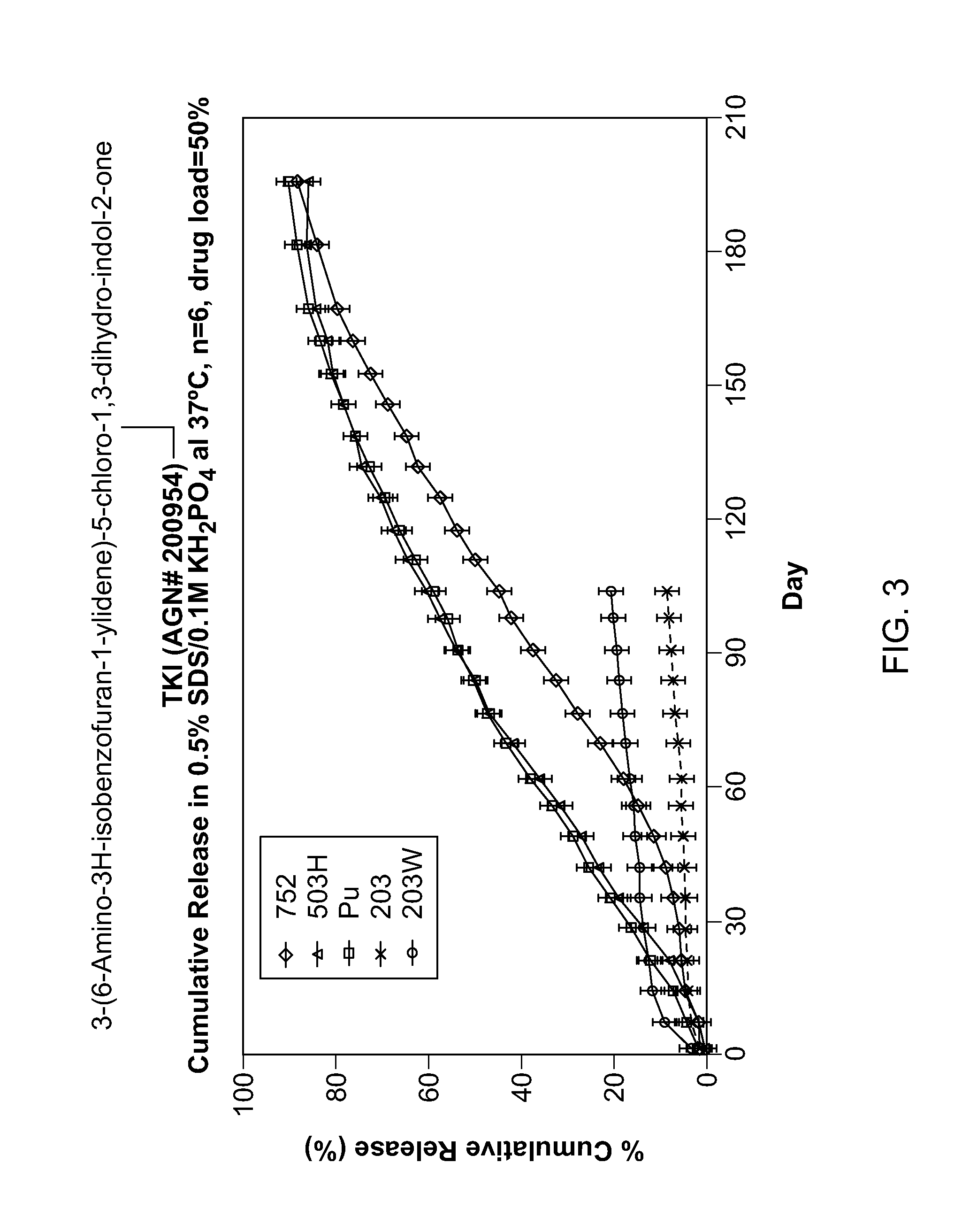
[0153]Tyrosine kinase inhibitors were incorporated into PLGA or PLA implants by extrusion. The TKIs were milled with the polymers at certain ratios then extruded into filaments. These filaments were subsequently cut into implants weighing approximately 1 mg. Several TKIs were formulated in the PLGA and PLA implants based on their potencies and physicochemical properties as shown in Table 2.

TABLE 2Tyrosine Kinase Inhibitors Formulated in PLGA ImplantsProjectedSolubilityAGNCss(μg / mL)NumberStructureEfficacypH 7log PpKaAGN 200954 4 ng / mL0.32.21 4.24 10.03AGN 202314 96 ng / mL2023.80 9.68AGN 202560105 ng / mL413.66 9.65AGN 201634 28 ng / mL881.25 4.06 10.25

[0154]TKI release from the implants was assessed in vitro. Implants were placed into vials containing release medium and shaken at 37° C. At the appropriate time points a sample was taken from the release medium for analysis and the medium totally replaced to maintain sink conditions. Drug in the sample was assayed ...

example 3

In Vivo Pharmacokinetic Properties of TKI-Containing Implants

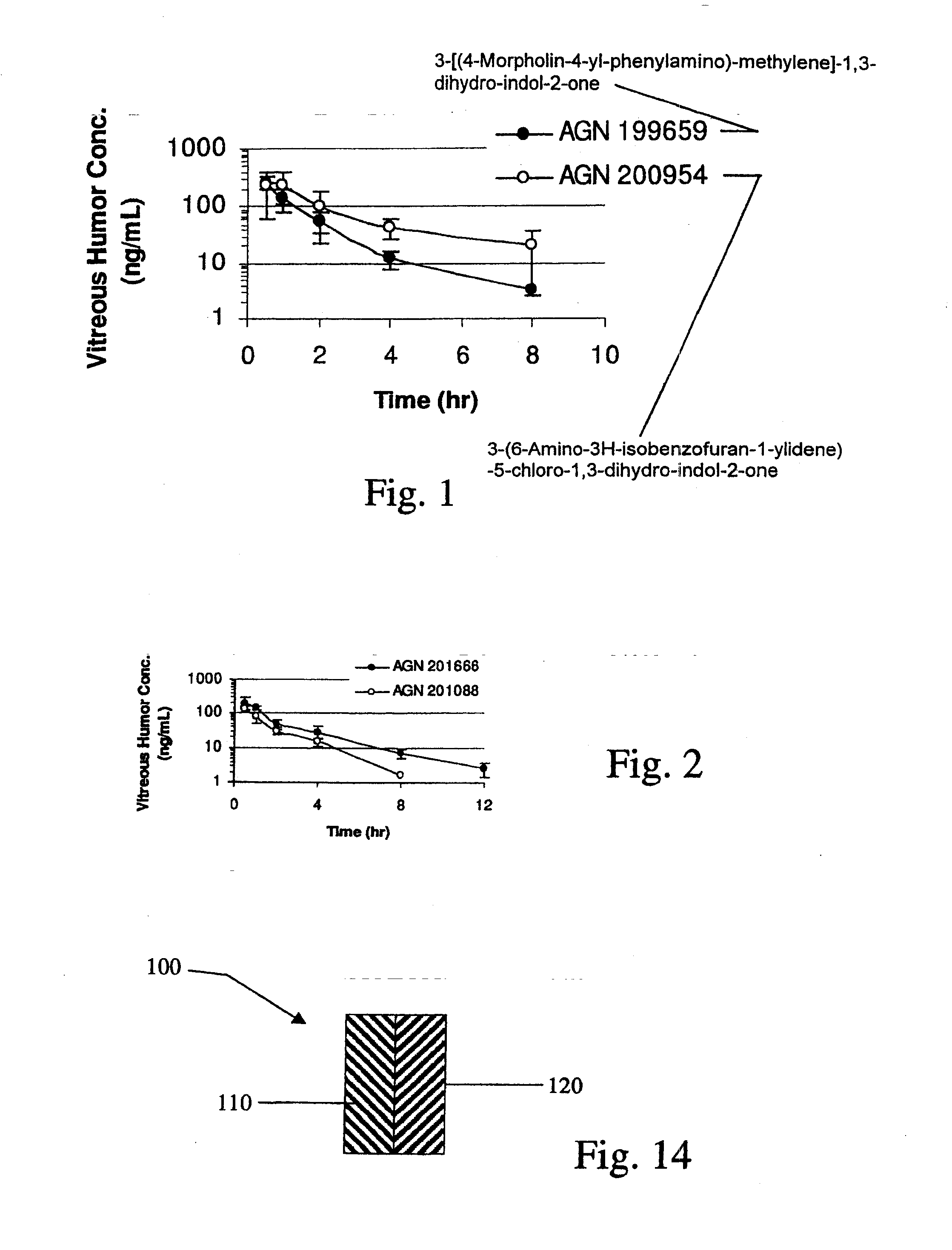
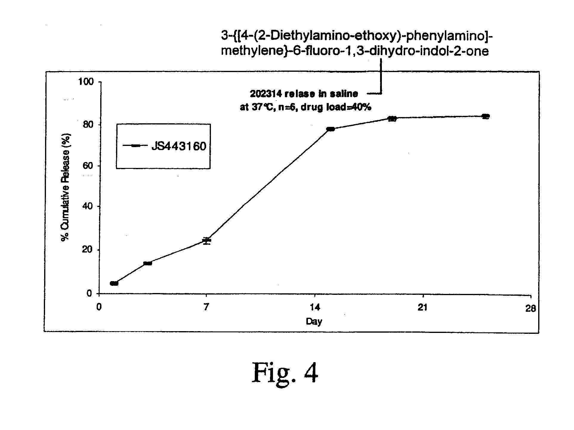
[0156]Implants containing AGN 202314 were placed intravitreally or subconjunctivally in an eye. The implants released AGN 202314 in-vitro over a 14 day period (FIG. 3.). The intent of this study was to achieve an intravitreal in-vivo / in-vitro correlation with the intravitreal implants and assess the feasibility of periocular delivery.

[0157]Intravitreal Implants, PLGA (400 μg AGN 202314 dose, 1 mg total implant weight), were implanted by surgical incision into the mid vitreous of albino rabbits. At days 8, 15, 31 and 61 rabbits were sacrificed and the vitreous humor, lens, aqueous humor and plasma assayed for AGN 202314.

[0158]Subconjunctival implants, PLGA (1200 μg AGN 202314 dose; three implants) and PLA microspheres (300 μg AGN 202314) were implanted subconjunctivally. At days 8, 15, 31 and 61 rabbits were sacrificed and the vitreous humor, lens, aqueous humor and plasma assayed for AGN 202314.

[0159]The data are summarize...

example 4

[0167]In vitro release of a TKI (AGN 201634) from an implant TKI release was examined for implants made from poly (D,L-lactide-co-glycolide) (PDLG) or poly (D,L-lactide) (PDL) in different media with or without addition of detergent at 37° C. in a shaking water bath.

[0168]AGN 201634 was obtained from Allergan, and its chemical structure is shown below. It was used as received without further purification. PDLG / PDL polymer materials were obtained from Purac America Inc.

[0169]TKI release was examined in various medium, including saline, phosphate buffer saline of pH 7.4, 50 mM bicarbonate buffer of pH 6.0±0.1 with 0.1% cetyltrimethylammonium bromide (CTAB), and 50 mM borate buffer of pH 8.5±0.1 with 0.5% sodium dodecyl sulfate (SDS) in a shaking water bath (Precision) at 37° C. Sample was incubated in 10 mL of medium, and was totally replaced with fresh medium at each sampling time. Drug concentration was determined by HPLC using a Waters 2690 Separation Module equipped with a Waters ...


PUM

PropertyMeasurementUnit
timeaaaaaaaaaa
timeaaaaaaaaaa
timeaaaaaaaaaa

Abstract

Biocompatible intraocular implants include a tyrosine kinase inhibitor and a biodegradable polymer that is effective to facilitate release of the tyrosine kinase inhibitor into the vitreous of an eye for an extended period of time. The therapeutic agents of the implants may be associated with a biodegradable polymer matrix, such as a matrix that is substantially free of a polyvinyl alcohol. The implants can be placed in an eye to treat or reduce the occurrence of one or more ocular conditions.

Description

CROSS REFERENCE[0001]This application is a divisional of copending U.S. patent application Ser. No. 11 / 119,001 filed Apr. 29, 2005, which is a continuation in part of U.S. patent application Ser. No. 10 / 837,361 filed Apr. 30, 2004, now issued as U.S. Pat. No. 7,771,742, the entire content of which are incorporated herein by reference.BACKGROUND[0002]The present invention generally relates to devices and methods to treat an eye of a patient, and more specifically to intraocular implants that provide extended release of a therapeutic agent to an eye in which the implant is placed, and to methods of making and using such implants, for example, to treat or reduce one or more symptoms of an ocular condition.[0003]Delivery of drugs to the retina, vitreous and uveal tract is typically achieved by high systemic dosing, intra-ocular injections or other heroic measures. Penetration of systemically administered drugs into the retina is severely restricted by the blood-retinal barriers (BRB) fo...

Claims


Application Information

Patent Timeline
Patent Type & Authority Applications(United States)
IPC IPC(8): A61K9/00A61K31/404A61F2/00A61K9/20A61K31/00
CPCA61K31/404A61K9/0051A61K9/204A61K31/454A61K31/5377A61P27/02A61F9/0017A61K47/34
Inventor EDELMAN, JEFFREY L.HUGHES, PATRICK M.MALONE, THOMAS C.DEVRIES, GERALD W.CHANG-LIN, JOAN-ENSHIAH, JANE-GUONIVAGGIOLI, THIERRYSPADA, LON T.BLANDA, WENDY M.
Owner ALLERGAN INC
Why Eureka
Eureka Blog
Learn More