
Decellularized small particle tissue

a small particle and tissue technology, applied in the field of decellularization and tissue production, can solve the problems of difficult to produce small particle tissue, unsatisfactory decellularized tissues in one way, and inability to retain debris and cellular materials, etc., to speed up the decellularization process, less manipulation of reagents, and high cost

Inactive Publication Date: 2013-10-31
BURKE EDMUND
View PDF2 Cites 4 Cited by
  • Summary
  • Abstract
  • Description
  • Claims
  • Application Information

AI Technical Summary

Benefits of technology

The present invention is about a technique for making decellularized tissue by using a variety of tissue sources, such as animal or human placenta, umbilical cord, skin, liver, kidney, spleen, and blood. The technique involves removing blood from the tissue and using a decellularizing agent to remove cells while keeping the tissue's structure intact. The resulting tissue can be further used in various applications, such as regenerative medicine or tissue engineering. The invention is advantageous in that it does not rely on a specific decellularizing agent and utilizes a sequential particle size separation and recycling system for the decellularization and particle size reduction steps, resulting in more efficient and cost-effective production of decellularized tissue.

Problems solved by technology

A major difficulty with most of the wide variety of processes and techniques that have been used to decellularize tissue, has been achieving the removal of the cells from the tissue while maintaining in the tissue, beneficial proteins from the starting materials which may be needed in the subsequent use of the decellularized tissue in its medical environment.
Similarly, while it is desirable, and in some cases necessary, to retain beneficial proteins, it is also undesirable to retain debris and cellular materials resulting from the decellularizing process since they may cause deleterious effects upon subsequent implantation into the body.
Nevertheless, it has been difficult to produce a small particle tissue while avoiding the difficulties expressed above.
These methods often yield decellularized tissues which are usually unsatisfactory in one way or another, however.
Other techniques involve methods which use harsh materials such as sodium hydroxide or hydrochloric acid under harsh conditions such as extremes in pH, without yielding generally suitable results.
Most of these methods are generally unwieldy requiring much manipulation in batch type processes and many of them, especially non-oxidizing acid extractions are not effective decellularizing agents and beneficial proteins remain unextracted to an unsuitable extent.
In short, it is felt that the present state of the prior art, while sufficient in some cases to remove some cellular materials, leaves much to be desired in terms of the quality of the decellularized tissue, especially in the loss of beneficial proteins remaining after the treatment with the decellularizing agents.

Method used


Image

  • Decellularized small particle tissue
  • Decellularized small particle tissue
  • Decellularized small particle tissue

Examples


example 1

[0078]This Example sets forth the materials and methods used in Examples 2-12.

Hydrogen peroxide used was from Acros Orrganics

2N NaOH and 2N HCL were from Fisher Scientific

Homegenizer used was a OMNI brand model GLH

Endotoxin analysis was done by USP method

Samples for protein analysis were digested in a pepsinacetic acid solution.

Collagen content was determined by Sirius Red method from Chondrex, Inc.

Fibronectin content was determined by the QuantiMatrix human fibronectin ELISA kit from Milipore.

Laminin content was determined by the QuantiMatrix human fibronectin ELISA kit from Milipore.

Glycosaminoglycan content was determined by the Blyscan Assay from Biocolor inc.

Elastin content was determined by the Fastin Assay from Biocolor, inc.

example 2

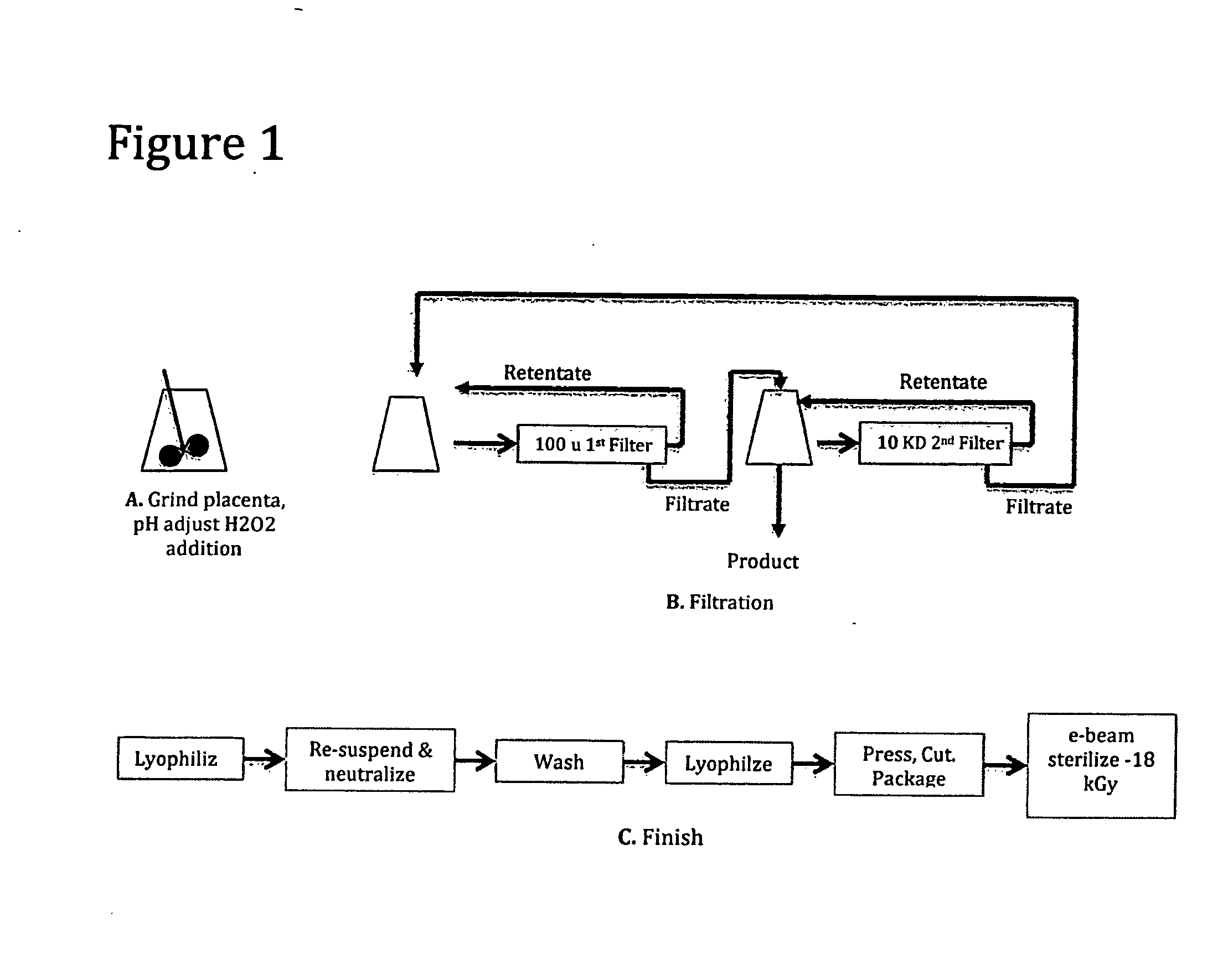
[0079]The following Example demonstrates a method of making decellularized small particle tissue from human placenta. A schematic flow of this method can be found in FIG. 1.

Human placenta including the attached umbilical cord and attached amnion and chorionic membrane were obtained from a normal birth. The tissue was obtained after blood was allowed to gravity drain from the cord into a bag for separate purposes. The placenta tissue was stored at −72 degrees C. until all appropriate viral testing and donor history could be reviewed by a qualified medical director in order that it could be released for research and development.

Grinding, pH Adjust and H2O2 Addition

[0080]772 grams of tissue was thawed overnight in a refrigerator and brought to room temperature on the day of processing. It was cut into small pieces (<3 cm in diameter), re-suspended in approximately 1.2 liter sterile water and then homogenized with a hand held homegenizer. A sample of untreated tissue was taken at this t...

example 3

[0085]A human placenta was processed similar to the decellularization and particle size reduction process of Example 2 and FIG. 1 except for the following changes.

The weight of the placenta on removal from the freezer was 465 grams.

The cut up placenta was homogenized with a Waring type blender.

The solids content of the ground unprocessed tissue material was 5.2%.

After the H202 addition, the slurry was mixed for an hour prior to filtration.

After filtration, approximately 85% of the starting material was captured as 2nd filter retentate and approximately 15% of the starting material was captured by as first filter retentate.

The filter used in finish processing step to wash the product had a 10 KD pore size.

[0086]The resulting product had the following characteristics:

The collagen content of the product as a percentage of dry material was equal to approximately 53% which is equal to 63% of the collagen content of pre-processed thawed and ground placenta as a percentage of dry material....


PUM

PropertyMeasurementUnit
pore sizesaaaaaaaaaa
pore sizeaaaaaaaaaa
particle sizeaaaaaaaaaa

Abstract

A process for producing a decellularized small particle tissue which involves selecting an appropriate tissue starting material from which a decellularized small particle tissue is desired to be prepared, treating the tissue with a decellularizing agent at an acid pH to remove at least a portion of the cellular material therefrom and to yield a product comprising a liquid component and a solid component, subjecting the liquid component and the solid component to a plurality of filters, F1 through Fn, wherein n may be 2, or an integer higher than 2, wherein the pore sizes of the filter F1 through Fn range from 200 microns to 10 Kilo Daltons, yielding filtrates and retentates, recycling either of said filtrates or said retentates or both, either separately or together, to any of steps b) or c) or both, at least one time, and isolating a decellularized small particle tissue from any of steps of the process. Both the product and process are novel.

Description

CROSS REFERENCE TO RELATED APPLICATIONS[0001]This application is a continuation-in-part of U.S. application Ser. No. 13 / 694,586 filed Dec. 14, 2012 which claims the benefit of U.S. Provisional Application No. 61 / 630,561 filed Dec. 14, 2011.STATEMENT REGARDING FEDERALLY SPONSORED RESEARCH OR DEVELOPMENT[0002](Not Applicable)REFERENCE TO A SEQUENCE LISTING A TABLE, OR A COMPUTER PROGRAM LISTING APPENDIX SUBMITTED ON A, COMPACT DISC (SEE 37 CFR 1.52(e)(5)[0003](Not Applicable)FIELD OF THE INVENTION[0004]The technical field to which this invention relates is the field of producing and decellularizing tissue. The art abounds with numerous techniques for producing decellularized tissues which are widely useable in such areas as tissue repair, tissue regeneration, wound repair, cell growth media or substrates, filling skin defects and voids, tissue implantation, skin grafting and regrowth, organ repair and organ transplantation and other similar areas.BACKGROUND OF INVENTION[0005]The numer...

Claims


Application Information

Patent Timeline
Patent Type & Authority Applications(United States)
IPC IPC(8): A61K35/12
CPCA61K35/12C12N2509/00C12N5/0605A61L27/26A61L27/3604A61L27/3687A61L27/3691A61L27/54A61L2300/414A61L2430/34A61L2430/40
Inventor BURKE, EDMUND
Owner BURKE EDMUND
Learn More