
Assay for Prions

a technology for prions and assays, applied in the field of assays for prions, can solve the problems of inability to accurately risk-assess the risk of future transfusion of vcjd, the current risk reduction strategy in the uk is extremely expensive, and significant problems for us public and military blood transfusion services, so as to avoid protease treatment, avoid immunoprecipitation steps, and excellent results

Inactive Publication Date: 2013-08-01
D GEN
View PDF14 Cites 32 Cited by
  • Summary
  • Abstract
  • Description
  • Claims
  • Application Information

AI Technical Summary

Benefits of technology

The invention relates to a method for detecting abnormal PrP in a sample of blood or urine, without the need for protease treatment or immunoprecipitation steps. The sample can be collected from a mammal, including livestock animals or humans. Dilution of the sample is crucial for signal generation, and optimal dilution ranges are provided for different species. The method can be used in preliminary steps of the invention, without the need for the subject to be present. The technical effect of the invention is a reliable and accurate method for detecting abnormal PrP in bodily samples.

Problems solved by technology

As the clinically silent incubation period of prion infection in humans can be prolonged, potentially exceeding 50 years (12), and the prevalence is poorly defined, the extent of future transfusion-transmission of vCJD cannot be estimated or accurately risk-assessed.
However, prior art assays typically assess infectivity in an animal model or cell culture system which is labour intensive and time consuming as well as raising ethical issues about use of animals.
Current risk reduction strategies in the UK are extremely costly involving leucodepletion of transfused packed red cells and the sourcing of plasma from the USA based on estimates of approximately 40% of infectivity being associated with leucocytes with the remainder in plasma (37).
These and related issues also apply outside the UK, for example blood donation is not permitted in the USA if the prospective donor has spent a cumulative period of 6 moths or more in Europe.
This causes significant issues for US public and military blood transfusion services.
It remains a major problem that a non-invasive and specific test for prion infection is not available.

Method used


Image

  • Assay for Prions
  • Assay for Prions
  • Assay for Prions

Examples


example 1

Detection of vCJD Brain Homogenate Spiked into Whole Blood

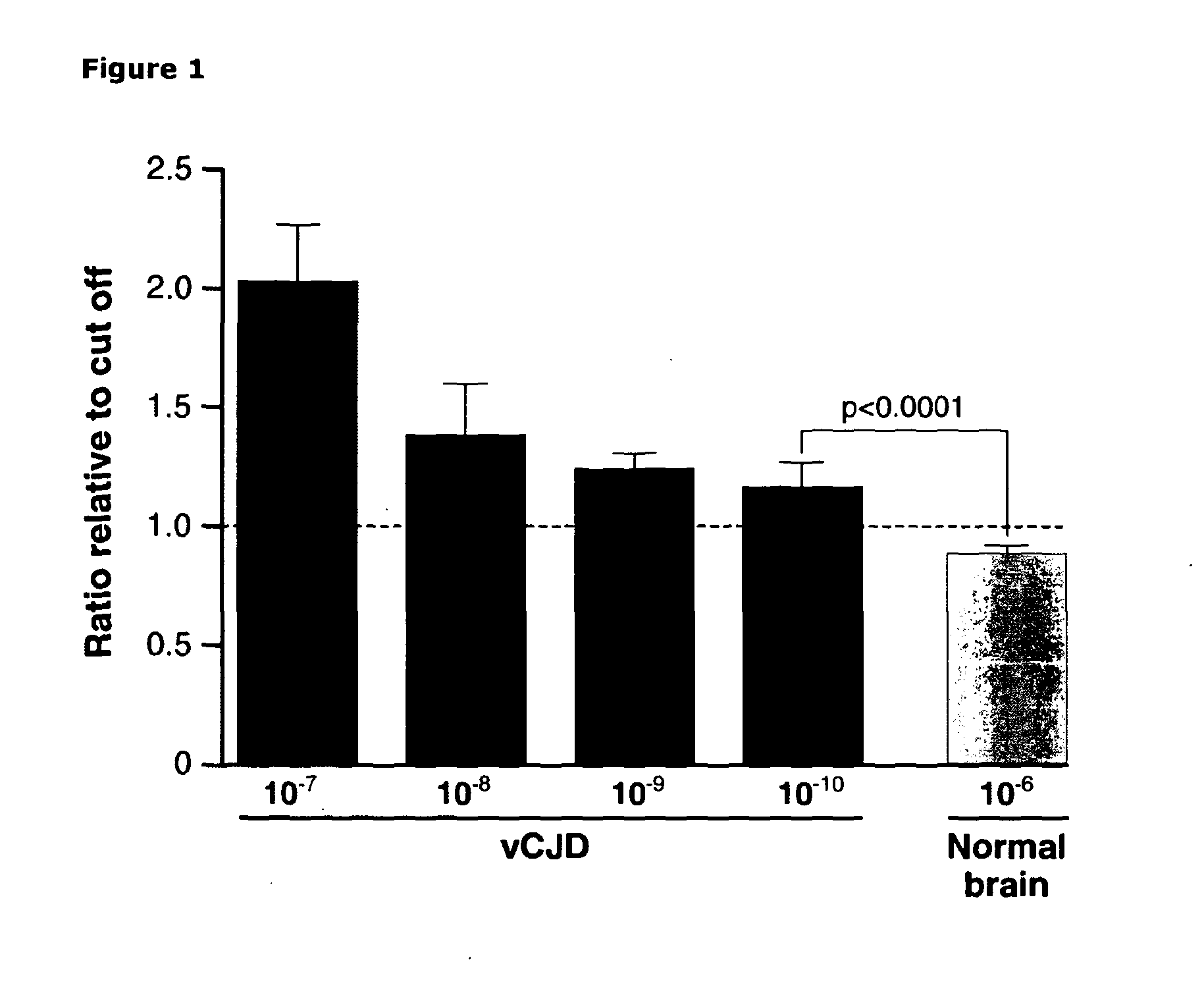
[0292]In order to determine the sensitivity of the assay relative to other methods we analysed serial dilutions of vCJD brain homogenate diluted into whole blood to provide a background diluent as close to endogenous patient samples as possible. A dilution range of 10−7 to 10−10 of 10% w / v vCJD brain homogenate was assayed and compared to a high background concentration (10−6) of normal brain homogenate (10% w / v) also diluted in whole blood. Although a non-linear response was seen with respect to dilution, vCJD-infected brain homogenate could clearly be distinguished from control even at a 1010 fold dilution (FIG. 1), a sensitivity more than 4 logs higher than previously achieved for immunoassay of vCJD tissue (31). A chemiluminescent signal of 1.3×10⁵+ / −1.1×10⁴ (mean+ / −SD) was obtained with 10−10 dilution of vCJD-infected brain versus 9.9×10⁴+ / −4.5×10³ for a 10−6 dilution of normal control brain. Data is expressed as a ratio...

example 2

Identification of vCJD Infected Patient Bloods

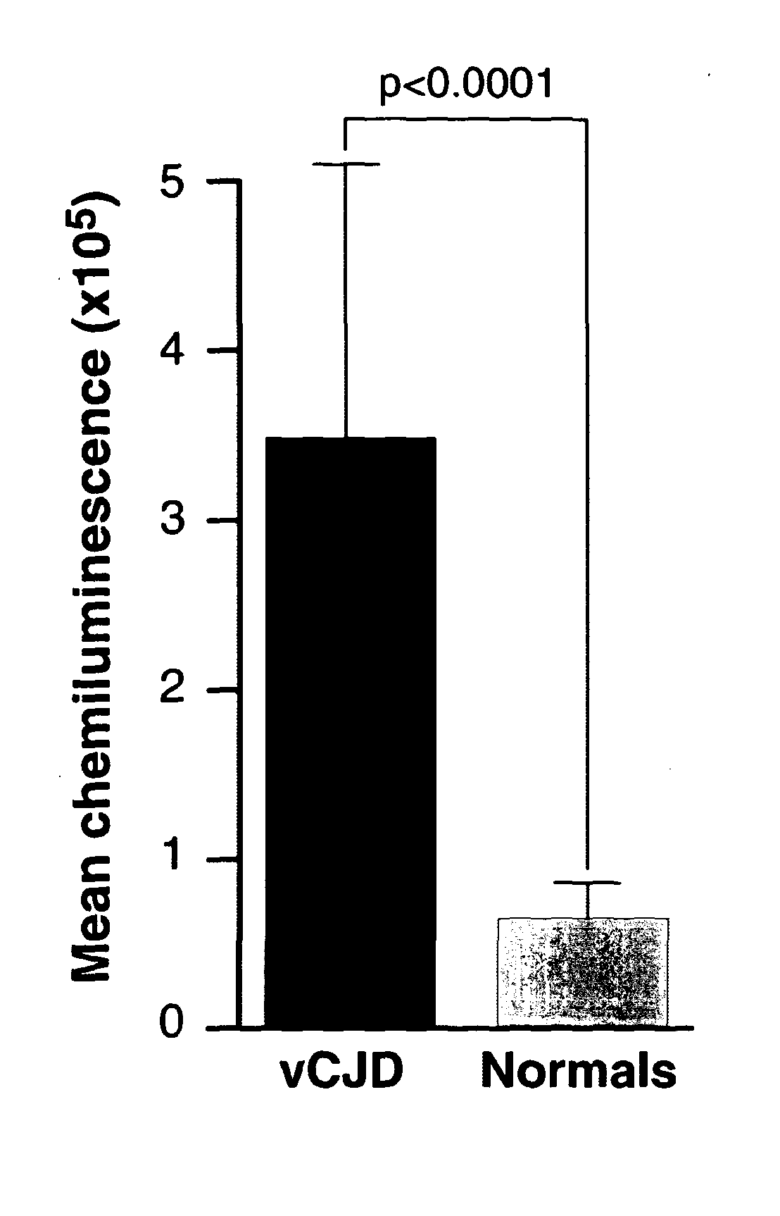
[0293]Initial studies performed using exogenous spikes of vCJD-infected brain homogenate in whole human blood demonstrated our assay was capable of discriminating between infected and non-infected samples with a sensitivity theoretically sufficient to detect infection in vCJD blood based upon estimates of titre obtained from rodent models (22;23). However, the biochemical nature of infectivity and abnormal PrP associated with blood is unknown and the results obtained from exogenous spiking experiments cannot be assumed to apply to authentic patient samples. To ensure this level of discrimination could be achieved with endogenous blood samples we tested a sub-set of confirmed vCJD patient bloods obtained from the National Prion Clinic and compared these to normal control bloods obtained from the NBS (FIG. 2). The samples were analysed as groups which had mean chemiluminescent signals that were significantly different (p value<0.0001, unpa...

example 3

Blind Panel Analysis

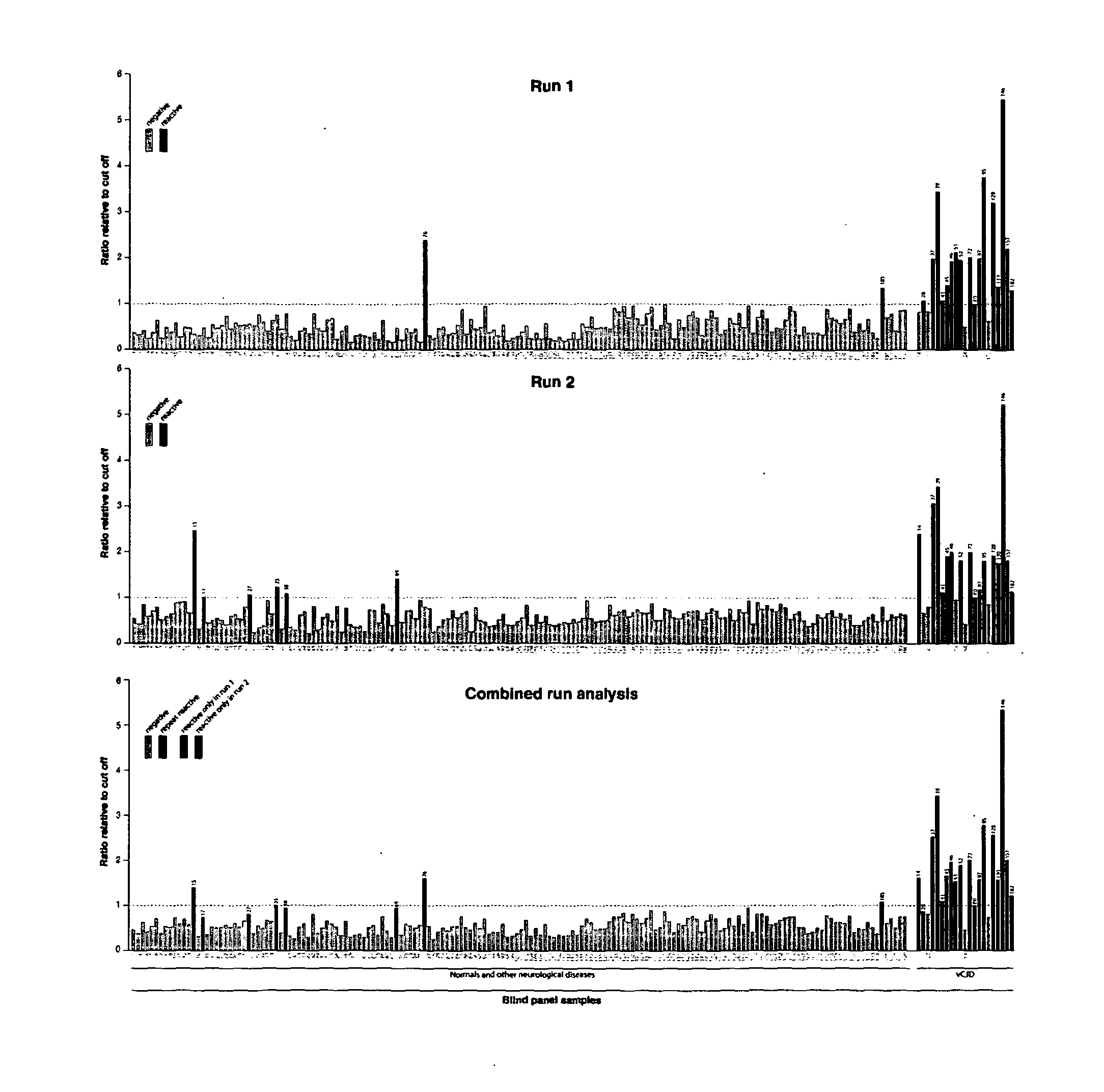
[0294]In order to confirm our results and remove any bias from the analysis a panel of 190 samples taken from 21 confirmed vCJD patients (National Prion Clinic), 69 patients with other neurological disease and 100 normal healthy controls (NBS) were blinded by parties independent to the testing and analysis. The samples were tested in batches of 19 samples with internal controls to determine a cut-off threshold for each plate, samples which gave chemiluminescence signals above the cut-off were deemed reactive. To eliminate potential false positive reactions originating not from the sample but from contamination or assay errors each sample was tested twice within independent assay runs (FIG. 3). Only those samples which were repeat reactive in both assays were scored as positive.

[0295]From the panel of 190 samples tested a total of 19 and 22 were reactive in assays 1 and 2 respectively (FIG. 3). A subset of 15 of those samples gave signals above the cut-off thresho...


PUM

PropertyMeasurementUnit
volumeaaaaaaaaaa
volumeaaaaaaaaaa
concentrationaaaaaaaaaa

Abstract

The invention relates to a method for detection of abnormal PrP in a sample of blood or urine, said method comprising: (a) diluting the sample with buffer to comprise final concentrations of (i) 10 mM to 500 mM buffer agent; (ii) 1% to 10% w / v bovine serum albumin; and (iii) 1% to 8% w / v CHAPS; (b) adding steel particles and incubating to allow PrP binding; (c) washing the steel particles to remove diluted sample; and (d) detecting abnormal PrP captured on the steel particles using antibody capable of binding said abnormal PrP. The invention also provides compositions and kits.

Description

FIELD OF THE INVENTION[0001]The invention relates to an assay for detecting prions. In particular the invention relates to an assay for detecting prion infection in blood or urine, most suitably blood.BACKGROUND TO THE INVENTION[0002]Prion diseases are a group of fatal neurodegenerative disorders including variant Creutzfeldt-Jakob disease (vCJD) which originates from exposure to bovine spongiform encephalopathy (BSE). Iatrogenic transmission from asymptomatic patients with vCJD prion infection via blood transfusion has had a major impact on public health policy.[0003]Prion Disease encompasses a range of closely related and uniformly fatal neurodegenerative disorders affecting the central nervous system of humans and animals. They include Creutzfeldt-Jakob disease (CJD), Gerstmann-Straussler-Scheinker disease (GSS), fatal familial insomnia (FFI) and kuru in humans, bovine spongiform encephalopathy (BSE) in cattle, chronic wasting disease (CWD) of deer and Elk and scrapie in sheep (1...

Claims


Application Information

Patent Timeline
Patent Type & Authority Applications(United States)
IPC IPC(8): G01N33/68
CPCG01N33/6896G01N33/6893G01N2800/2828G01N2333/70596
Inventor JACKSON, GRAHAM STUARTCOLLINGE, JOHNEDGEWORTH, JULIE ANN
Owner D GEN
Learn More