Single molecule spectroscopy for analysis of cell-free nucleic acid biomarkers

a single molecule, nucleic acid technology, applied in fluorescence/phosphorescence, instruments, laboratory glassware, etc., can solve the problems of rapid and efficient translation, high cost of pcr-based diagnostic assays, laborious, time-consuming,

Inactive Publication Date: 2012-05-31
THE JOHN HOPKINS UNIV SCHOOL OF MEDICINE
View PDF3 Cites 57 Cited by
  • Summary
  • Abstract
  • Description
  • Claims
  • Application Information

AI Technical Summary

Benefits of technology

[0251]An embodiment of the current invention is directed to a microfluidic device that includes inline micro-evaporators to concentrate biological target molecules within nano-to-picoliter-sized water-in-oil droplets. These droplets can serve as both low-volume reactors for parallel sample processing of the concentrated samples, and digital compartments that enable ordered transfer for downstream SMD analysis. Utilization of the evaporators as microliter-to-picoliter interconnects between the macroscopic world and single molecule microanalytical systems can solve problems of conventional devices such as those discussed above that hinder the widespread acceptance and utilization of SMD. First, solvent removal within the evaporators transports and confines the molecular contents of large sample volumes to the downstream droplets, which can be swept through laser-illuminated, confocal fluorescence detection volumes. The intradroplet, molecular detection efficiency at this point can be as high as about 100% using cylindrical illumination confocal spectroscopy (CICS) (K. H. Liu and T. H. Wang. Biophys. Journal, 95(6), 2964-2975, 2008) and pushing the entire droplet through a laser-illuminated sheet; however, the optical probe can be made to match a variety of operational parameters and the platform is not limited to only CICS detection. Unlike traditional continuous flow SMD platforms, sample throughput and the kinetics of probe-target interactions of single molecule assays conducted in accordance with some embodiments of the current invention are limited by the speed of solvent removal, which is a controllable device parameter. Therefore, run times for single molecule assays can be greatly reduced due to target enrichment within the droplets, which facilitates probe-target interactions at relatively high concentrations. At these concentrations, droplet-based microfluidics becomes an advantageous complementary technology to single molecule optical platforms, allowing rapid analysis of molecules trapped within parallel reaction compartments in an automated and controllable fashion. And, in addition to simply making SMD amenable to high-throughput studies of genetic alterations, microfluidic systems and methods according to some embodiments of the current invention can open new biological applications that were previously unachievable. For instance, microfluidic loading and quick analytical schemes according to some aspects of the current invention can make high-speed, fluorescence-activated molecular sorting (“FACS for molecules”) a possibility, within controllable reaction compartments that can be manipulated and observed nearly at the will of the genomic researcher.
[0315]Microevaporators could easily be integrated with other detection schemes, such as disk and wire-like nano-biosensors (Z. Gao, A. Agarwal, A. D. Trigg, N. Singh, C. Fang, C. H. Tung, Y. Fan, K. D. Buddharaju and J. Kong, Anal. Chem., 2007, 79, 3291-3291-3297; F. Patolsky, G. Zheng and C. M. Lieber, Nanomed., 2006, 1, 51-51-65) to increase analyte transfer and kinetics of target capture. Detection chambers for these nanoscale biosensors could reach picoliter levels, enabling concentration factors surpassing the ˜6500 shown using nanoliter chambers in this example. Indeed, optimization and standardization of microevaporators as universal analyte inputs to microanalytical systems could lift many of the current limitations of conventional microfluidic delivery systems. Additional improvements to membrane-based evaporators could include ion permeable membranes, enabling control over buffer concentrations during solvent removal, thus expanding applicability to complex protein and microorganism containing samples. Further modifications to the evaporator coil could also include the use of three-dimensional microstructures to maximize the surface area of the pervaporation membrane, which would lead to increases in assay sensitivity, while substantially decreasing total processing time. In this manner, processing times for single molecule detection platforms, such as single molecule fluorescence counting, that are traditionally limited due to probe-target hybridization kinetics would become dominated by the controllable evaporation or enrichment speeds within the evaporation-based analyte input. In addition, utilizing solvent removal as a simple method of analyte transport alleviates many of the challenges involved with low-volume sample processing and the lack of compatibility between conventional lab methodologies and SMD. Therefore, these results represent a clear example that for specific biological applications the performance of any microanalytical device must be assessed by the sensitivity of the sum of its parts, and not just the responsiveness of its probe.

Problems solved by technology

Unfortunately, PCR-based techniques are fraught with technical and practical limitations that have precluded the rapid and efficient translation of CNA biomarkers from the discovery stage into clinical practice.
For example, PCR-based diagnostic assays are expensive, labor intensive, time consuming, and difficult to reproduce on a daily basis.
In addition, PCR based assays cannot be easily multiplexed, limiting the number of markers that can be concurrently analyzed.
Finally, it is challenging to perform accurate quantification of low level changes in CNA biomarkers using PCR.
These limitations have hindered the clinical validation and adoption of these promising biomarker molecules.

Method used


Image

  • Single molecule spectroscopy for analysis of cell-free nucleic acid biomarkers
  • Single molecule spectroscopy for analysis of cell-free nucleic acid biomarkers
  • Single molecule spectroscopy for analysis of cell-free nucleic acid biomarkers

Examples


example i

Applications of a Method of the Invention

A. 1-Step CNA Analysis.

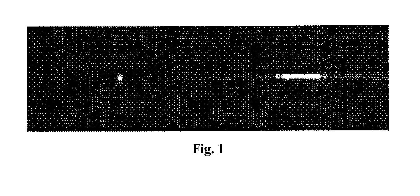
[0170]Microfluidic Cylindrical Illumination Confocal Spectroscopy (μCICS) is ideally suited for the clinical analysis of CNAs. In μCICS, the standard diffraction limited CS observation volume is elongated in 1D to span the entire microchannel as illustrated inFIG. 1. The 1D expansion increases the mass detection efficiency to 100% and greatly enhances the analysis uniformity. Thus, it increases throughput, enables more accurate determination of molecular properties, and enables assays that are impossible to efficiently perform using other methods.

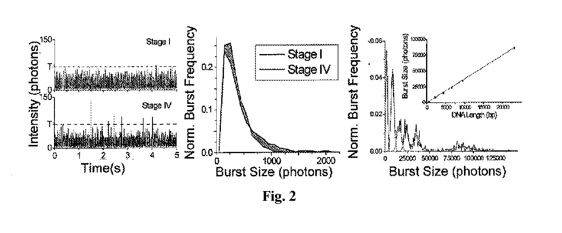
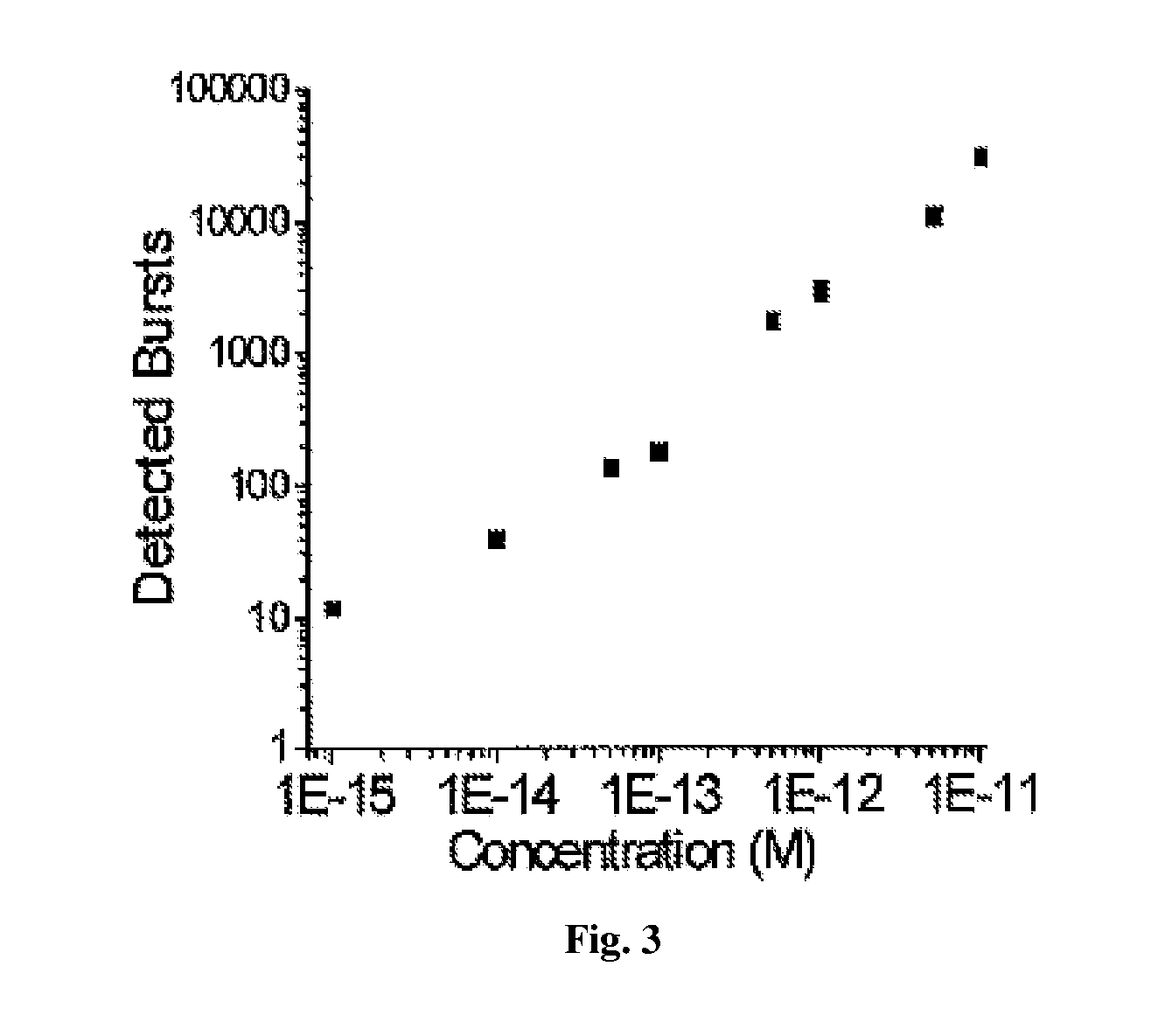
[0171]Using the μCICS platform, we have performed CNA analysis directly from serum with a 1-step assay called single molecule DNA integrity analysis (smDIA). With this assay, we are able to directly measure both DNA integrity (i.e. DNA fragment size) and DNA quantity without PCR amplification, DNA isolation, or separation steps. Previous studies have shown that the DNA integrit...

example ii

System for CICS

[0183]The terms light, optical, optics, etc are not intended to be limited to only visible light in the broader concepts. For example, they could include infrared and / or ultraviolet regions of the electromagnetic spectrum according to some embodiments of the current invention.

[0184]An embodiment of the current invention provides a confocal spectroscopy system that can enable highly quantitative, continuous flow, single molecule analysis with high uniformity and high mass detection efficiency. Such a system will be referred to as a Cylindrical Illumination Confocal Spectroscopy (CICS) system. CICS is designed to be a highly sensitive and high throughput detection method that can be generically integrated into microfluidic systems without additional microfluidic components.

[0185]Rather than use a minute, diffraction limited point, CICS uses a sheet-like observation volume that can substantially entirely span the cross-section of a microchannel. It is created through the...

example iii

Microfluidic System for High-throughput, Droplet-Based Single Molecule Analysis with Low Reagent Consumption

SUMMARY

[0247]A microfluidic device for a confocal fluorescence detection system according to an embodiment of the current invention has an input channel defined by a body of the microfluidic device, a sample concentration section defined by the body of the microfluidic device and in fluid connection with the input channel, a mixing section defined by the body of the microfluidic device and in fluid connection with the concentration section, and a detection region that is at least partially transparent to illumination light of the confocal fluorescence detection system and at least partially transparent to fluorescent light when emitted from a sample under observation as the sample flows through the detection region.

[0248]A microfluidic detection system according to an embodiment of the current invention has a microfluidic device having a detection region defined by a body of t...


PUM

PropertyMeasurementUnit
Volumeaaaaaaaaaa
Volumeaaaaaaaaaa
Volumeaaaaaaaaaa

Abstract

The present invention relates, e.g., to a method for detecting a nucleic acid molecule of interest in a sample comprising cell-free nucleic acids, comprising fluorescently labeling the nucleic acid molecule of interest, by specifically binding a fluorescently labeled nanosensor or probe to the nucleic acid of interest, or by enzymatically incorporating a fluorescent probe or dye into the nucleic acid of interest, illuminating the fluorescently labeled nucleic acid molecule, causing it to emit fluorescent light, and measuring the level of fluorescence by single molecule spectroscopy, wherein the detection of a fluorescent signal is indicative of the presence of the nucleic acid of interest in the sample.

Description

[0001]This application claims the benefit of the filing date of U.S. provisional application 61 / 176,745, filed May 8, 2009, which is incorporated by reference herein in its entirety.[0002]This research was supported by grants from NIH (1R21CA120742) and NSF (0725528 and 0552063). The U.S. government thus has certain rights in the invention.FIELD OF THE INVENTION[0003]This invention relates, e.g., to a diagnostic method for detecting biomarkers in single molecule cell-free nucleic acid, using single molecule spectroscopy.BACKGROUND INFORMATION[0004]Cell-free nucleic acids (CNAs) are a highly promising source of non-invasive biomarkers for the detection of a wide array of human diseases. CNAs are extra-cellular nucleic acids freely present in human body fluids such as blood, urine, and sputum. This makes them easily obtained and highly attractive as a source of non-invasive biomarkers. They are released by both diseased and healthy cells alike and have been used to diagnose and manage...

Claims


Application Information

Patent Timeline
IPC IPC(8): G01N21/64B01L3/00C40B30/04B82Y15/00
CPCC12Q1/6844C12Q2563/143C12Q2549/119C12Q2537/149
Inventor WANG, JEFF TZA-HUEILIU, KELVIN J.PULEO, CHRISTOPHER M.
Owner THE JOHN HOPKINS UNIV SCHOOL OF MEDICINE
Why Eureka
Try Eureka