Differentiation of Human Embryonic and Induced Pluripotent Stem Cells

Inactive Publication Date: 2011-09-22
UNIV OF CONNECTICUT
View PDF6 Cites 9 Cited by
  • Summary
  • Abstract
  • Description
  • Claims
  • Application Information

AI Technical Summary

Benefits of technology

[0005]The invention solves many of the problem associated with such earlier methods and is based, in part, on culture systems and conditions that promote the rapid, direct, progressive, and substantially uniform differentiation of stem cells such as undifferentiated pluripotent human embryonic stem cells (hESCs) or induced pluripotent stem cells (iPSCs or iPS cells) into the chondrogenic lineage without prior EB formation. Stem cells obtained from cord blood or amniotic fluid are also used in the methods. The substantially pure chondrogenic cells at defined stages of differentiation that are produced using the methods provide a solution to the problems and drawbacks of cell heterogeneity (with respect to tissue type as well as stage of differentiation) associated with prior methods. The cell populations described herein are substantially free of non-chondrogenic cells, e.g., the populations comprise at least 85%, 95-98%, and up to 99 or 100% chondrogenic cells. The invention also provides high density culture conditions and additional procedures which promote the progression of the differentiation of ESCs into the chondrogenic lineage and which contribute to the uniformity of chondrogenic differentiation achieved.
[0013]The culture systems and conditions of the invention avoid the cellular heterogeneity that complicates the use of cells derived from embryoid bodies (EBs) for cell-based cartilage repair therapies. Since the methods promote direct and uniform differentiation and circumvent the EB stage, they reliably yield cell populations at a defined stage of differentiation, i.e., early stage versus late stage. Gene expression profiling enables the identification of hESC-derived progenitor cells in different stages of the chondrogenic lineage, ranging from cells just entering into the chondrogenic lineage to overtly differentiated chondrocytes. In addition to continuing to differentiate, cells in earlier phases of the chondrogenic lineage (e.g., bearing early cartilage markers such as Sox9) respond to local microenvironmental signals in situ after they are transplanted into a recipient and are thus desirable for participation in cartilage repair compared to cells at late stages of the lineage. Cells in late stages of the chondrogenic lineage (e.g., bearing markers indicative of more overtly differentiated cells cartilage markers such as aggrecan) are suitable for cartilage repair / restoration procedures. Thus, unlike earlier methods that yielded a heterogeneous population of cells at various stages of differentiation and different lineages, the present methods provide a source of staged cells and permit fine tailoring of therapies based on the stage of differentiation best suited to the pathological condition to be treated.
[0021]A significant advantage of the methods is that they permit isolation of substantially pure populations of cells at defined stages of differentiation, e.g., at a stage where cells are just beginning to differentiate (e.g., day 2 cells, day 3 cells, or day 4 cells) or fully differentiated chondrocytes (e.g., day 7-14 cells). Fully differentiated chondrocytes are useful for numerous cartilage repair and restoration therapies. Cells at earlier stages of differentiation are used for the same purpose or for other purposes with the added advantage that they colonize and undergo further differentiation as well as respond to local environmental signals in situ.

Problems solved by technology

Treatment of degenerative cartilage diseases is a particular clinical challenge because of the limited capacity of the tissue for self-repair.
Although several culture systems have been developed in which ESC-derived cells differentiate to various degrees into chondrocytes, in these systems chondrogenic differentiation is not uniform, and chondrocytes represent only a subpopulation of the cells that differentiate, complicating utilization of the cell population for cartilage repair.
A drawback of such earlier methods is that the cellular heterogeneity of EB-derived cells hinders the ability to obtain homogeneous populations of chondrogenic cells that can be used for cartilage repair.

Method used


Image

  • Differentiation of Human Embryonic and Induced Pluripotent Stem Cells
  • Differentiation of Human Embryonic and Induced Pluripotent Stem Cells
  • Differentiation of Human Embryonic and Induced Pluripotent Stem Cells

Examples


example 1

Culture of hESCs and Embryoid Body Formation

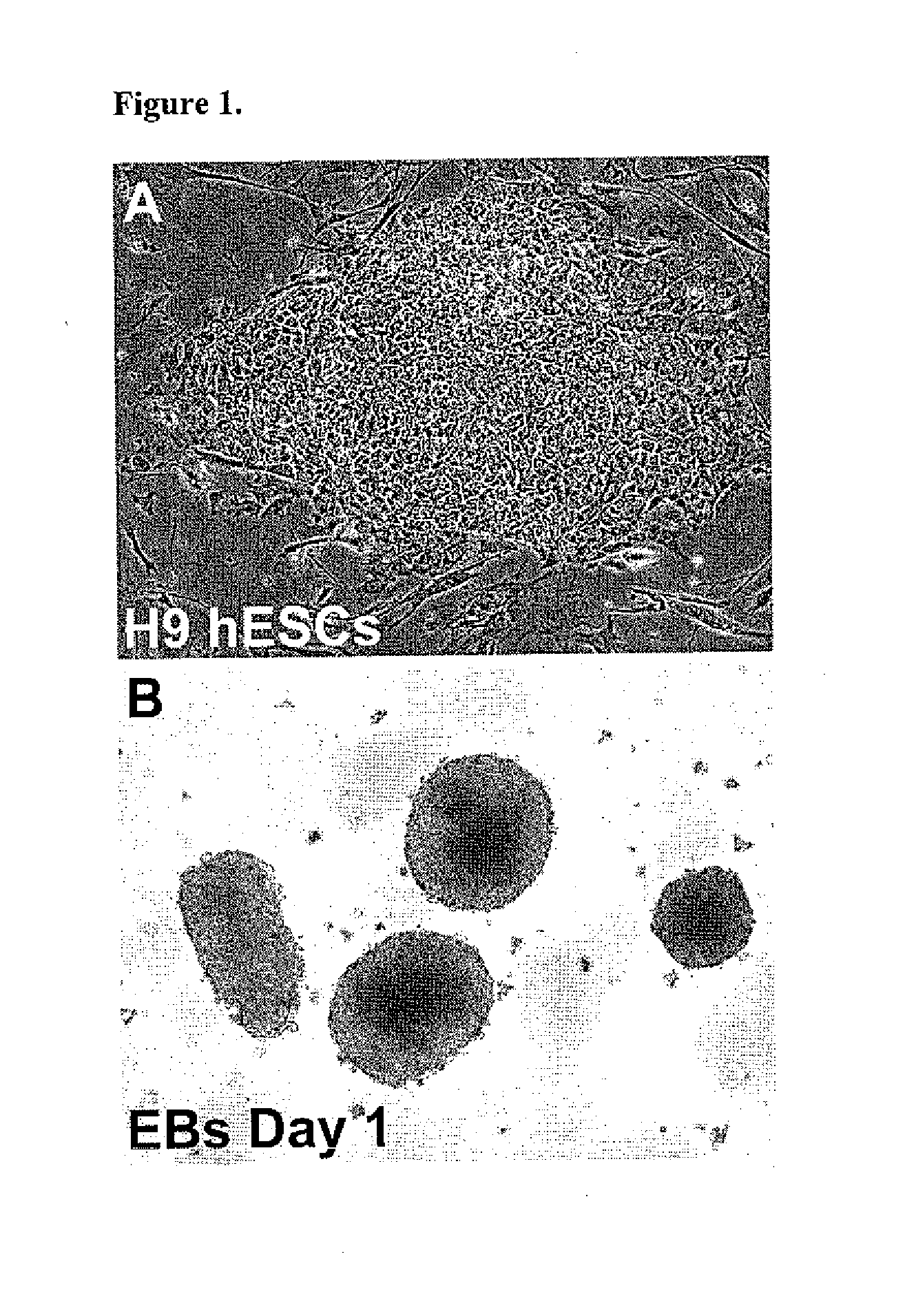
[0069]The H9 hESC line generated at the WiCell Research Institute (Thomson J A et al., 1998 Science, 282:1145-1147). The H9 cells were cultured in the serum-free hESC medium previously described (Toh W S et al., 2007 Stem Cells, 25:950-960) on a feeder layer of irradiated CF1 mouse embryonic fibroblasts (MEFs) (see, FIG. 1A) prepared using standard protocols by the University of Connecticut Stem Cell Core. Colonies were passaged every 4-6 days after treatment with 1 mg / ml collagenase IV (Invitrogen). hESCs from passage numbers 36 to 45 were used in these studies.

[0070]To generate EBs, hESC colonies were detached from the MEF feeder layers by treatment with 0.1% dispase (Invitrogen) for 20-30 minutes at 37° C., suspended in EB formation medium (Toh W S et al., 2007 Stem Cells, 25:950-960) in T75 Ultra-Low Attachment Flasks (Corning, Lowell, Mass.), and incubated for 5 days. Media was changed every 48 hours. The suspended hESC colonies form ...

example 2

Direct and Progressive Chondrogenic Differentiation of Undifferentiated Pluripotent hESCs

[0074]In view of the ill-defined cellular environments and inherent cellular heterogeneity characteristic of EBs, the ability of undifferentiated pluripotent H9 hESCs (FIG. 1A) to directly differentiate into the chondrogenic lineage when subjected to high density culture was examined. High density cultures are characterized by a concentration of greater than 1×10⁵ cells per 10 μl of medium. For example, the concentration of cells is in the range of 1-4×10⁵ cells per 10 μl of medium, e.g., 2×10⁵ cells per 10 μl. The volumes are scaled up as desired for larger scale cultures provided that the cell density / ratio remains in the range described above.

[0075]One day after their establishment, micromass cultures of dissociated H9 cells were supplied with serum-free “chondrogenic medium” in the presence and absence of 100 ng / ml of BMP2. As shown in FIG. 4A, only a few small Alcian blue staining areas wer...

example 3

Characterization of Cells in Various Phases of the Chondrogenic Lineage in hESC Micromass Cultures by Gene Expression Profiling

[0078]To confirm and quantify the progression of the differentiation of pluripotent hESCs into the chondrogenic lineage and to further characterize progenitor cells in different phases of the lineage, the expression of marker genes characteristic of different stages of the chondrogenic lineage was examined by quantitative Real Time RT-PCR. Gene expression patterns were analyzed at various times in BMP2-treated micromass cultures of pluripotent hESCs subjected to the modified chondrogenic differentiation protocol that leads to the progressive chondrogenic differentiation assayed by Alcian blue matrix staining as shown in FIGS. 5D-F above.

[0079]As shown in FIG. 7A, at the end of day 3 of culture, which is 24 hours after the addition of exogenous BMP2, there is a striking 7-fold upregulation in the expression of the chondrogenic transcription factor Sox9. Sox9 ...


PUM

PropertyMeasurementUnit
Fractionaaaaaaaaaa
Fractionaaaaaaaaaa
Timeaaaaaaaaaa

Abstract

The invention relates to culture systems, methods, and conditions that allow pluripotent undifferentiated hESCs or iPSCs to progressively and uniformly differentiate into cells of the chondrogenic lineage.

Description

RELATED APPLICATION INFORMATION[0001]This application claims priority to U.S. Provisional Patent Application Ser. No. 61 / 264,170, filed on Nov. 24, 2009.FIELD OF THE INVENTION[0002]The invention generally relates to methods for directing the differentiation of human embryonic and induced pluripotent stem cells into the cartilage lineage.BACKGROUND OF THE INVENTION[0003]Degenerative diseases of cartilage, e.g., osteoarthritis, are among the most prevalent and debilitating chronic health problems in the United States. Treatment of degenerative cartilage diseases is a particular clinical challenge because of the limited capacity of the tissue for self-repair. Because of their ability to differentiate into multiple cell types and their unlimited capacity for self-renewal, human embryonic stem cells (hESCs) are a potentially powerful tool for the repair of cartilage defects. Fulfilling the potential of hESCs for repair of diseased and damaged cartilage requires developing methods for dir...

Claims


Application Information

Patent Timeline
IPC IPC(8): A61K35/12C12N5/0735C12N5/0775C12N5/077C12Q1/02C12Q1/68A61P19/04A61P19/02A61P19/08A61P43/00
CPCC12N5/0655C12N2501/155C12N2506/45C12N2506/02A61P19/00A61P19/02A61P19/04A61P19/08A61P43/00
Inventor DEALY, CAROLINEKOSHER, ROBERT
Owner UNIV OF CONNECTICUT
Why Eureka
Try Eureka