
Biologic matrices comprising Anti-infective methods and compositions related thereto

a technology of biological matrices and compositions, applied in the direction of antibacterial agents, prostheses, ligaments, etc., can solve the problems of increasing the risk of infection for invasive medical devices, increasing the likelihood of opportunistic infection, and creating the risk of infection

Inactive Publication Date: 2011-03-24
MUSCULOSKELETAL TRANSPLANT FOUND INC
View PDF9 Cites 7 Cited by
  • Summary
  • Abstract
  • Description
  • Claims
  • Application Information

AI Technical Summary

Benefits of technology

The patent describes a method of treating an acellular biologic matrix with an anti-infective agent, such as triclosan. The matrix can be hydrated with a solution of specially denatured alcohol and then dipped in a solution containing the anti-infective agent. The treated matrix can also be vacuum-dried or sprayed with the anti-infective agent. The treated matrix has been found to have anti-microbial activity against gram positive bacteria and is safe for use in implantation procedures. The technical effect of this patent is to provide a biologic matrix with improved anti-infective properties for use in medical applications.

Problems solved by technology

Whenever a medical device is in contact with a patient, a risk of infection is created.
The risk of infection increases dramatically for invasive medical devices, such as intravenous catheters, arterial grafts, intrathecal or intracerebral shunts, and prosthetic devices, which are in intimate contact with bodily tissues and fluids.
Even under highly aseptic conditions, implantation requires surgical incisions in the subject, thereby increasing the likelihood of opportunistic infection.
Additionally, infection may have been present in the subject prior to surgery, or a wound may have been contaminated from a traumatic incident to the patient, or improper handling of the patch prior to its use may result in microbial contamination.
Synthetic or biologic mesh used in abdominal hernia repair, for example, may become infected in patients.
A serious problem with the use of a biologic mesh in infected ventral hernia applications is the tendency of the bacteria to cause the implant to be degraded and resorbed.

Method used


Image

  • Biologic matrices comprising Anti-infective methods and compositions related thereto

Examples


example 1

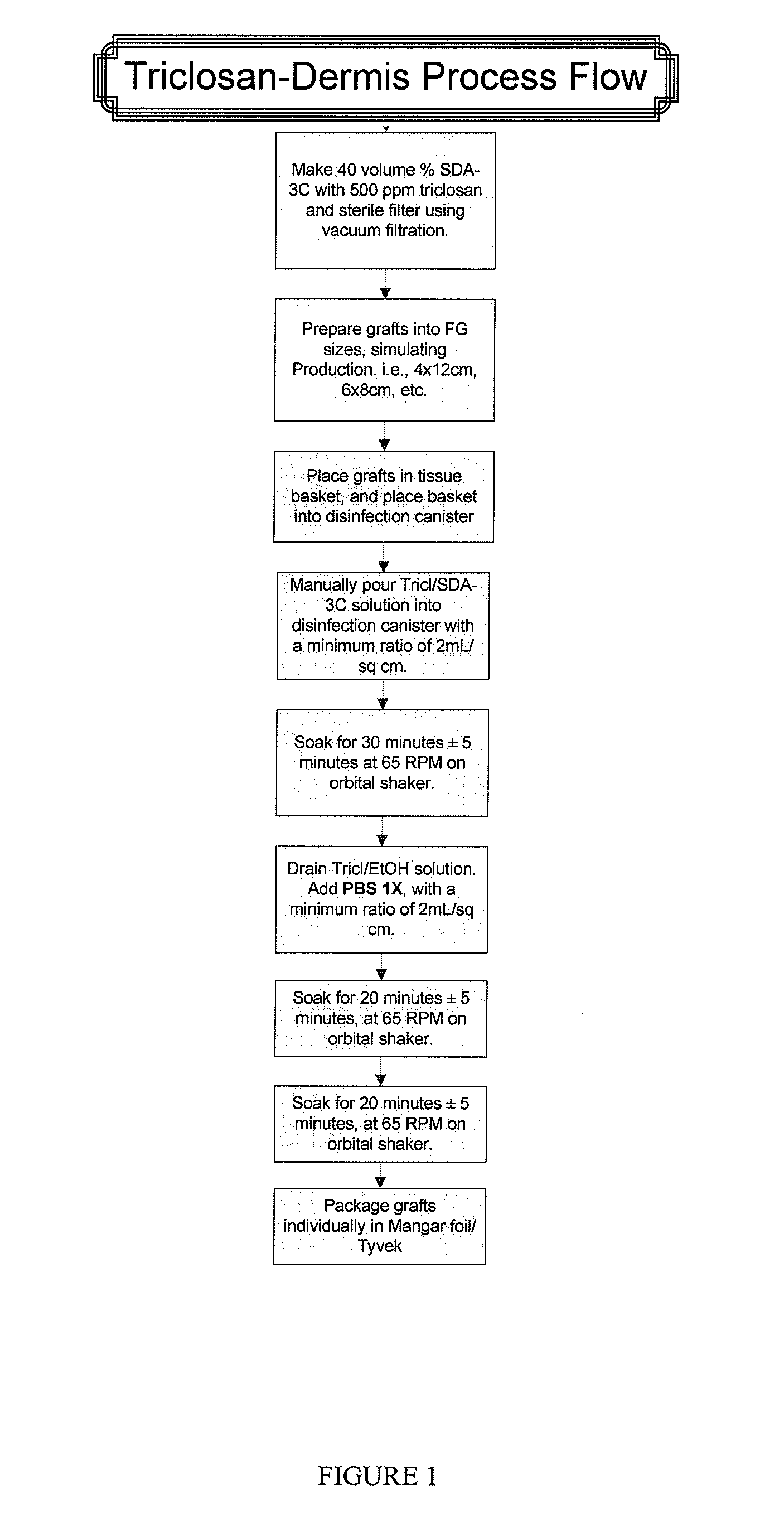
Sample Process for Dipping Acellular Dermis in a Triclosan Solution

[0070]A. Prepare sterile Triclosan in 70% SDA solution:

[0071]Aseptically mix 4000 ppm Triclosan in 140 proof ethanol on the bench top in a glass bottle. Pour contents of the bottle into the top of the filtration unit and filter.

B. Soak Dermis in physiologic salt solution:

[0072]The thickness of dermis should be less than or equal to 2.5 mm.

[0073]Fill container(s) with Dulbecco's balanced salt solution (Gibco) (Invitrogen 1404141).

[0074]Any container can be used to hold the salt solution. For example, a suitable container includes a plastic Nalgene container, which holds 600 mL of solution. Another suitable container is a metal pan, which holds 1000 mL of solution. There is a typical minimum ratio of solution to cm2 of dermis. The ratio is 0.69 mL salt solution / cm2 dermis. Typically, there is no maximum ratio.

[0075]Place graft between two sterile wipes and squeeze out excess liquid.

[0076]Submerge the graft in Dulbecco'...

example 2

Evaluation of Dermis with Anti-Infective by Inoculation and Log-Reduction Time-Course Survivor Counts

Method

[0084]Inoculation of the Test Material

[0085]A dilution of the 24 hours test organism to achieve a concentration of approximately 105 to 106 cfu per square samples approximately (1″×1″) was prepared in duplicate. 10 μL of test organism suspension was placed onto each square sample. Immediately after each time period (including an immediate recovery from product at zero-time) the inoculated sample was placed in Waring Blender containing 100 mL of GBL Stat Broth and macerated for 10-15 seconds. Recovery was performed by plate count as follows:

[0086]10 mL to give a dilution of 10−1 plated with GBL Stat Agar

[0087]1 mL to give a dilution of 10−2 plated with GBL Stat Agar

[0088]1 mL to 9.0 mL of saline and performed two fold serial dilution as 10−3 and 10−4 and 1.0 mL aliquots were plated with GBL Stat Agar.

[0089]The plates were incubated for 48 to 72 hours at 30 to 35° C. At the end o...

example 3

Evaluation of Dermis with Anti-Infective by Zone-of-Inhibition Assay

Inoculum Preparation

[0104]The Staphylococcus aureus (MRSA) was grown into 15 mL of Trypticase Soy Broth at 30 to 35 C for 18 to 24 hours and then diluted 1:1000 in sterile saline.

Procedure

[0105]The recovery media plates were surface-streaked with 0.5 mL of a 1:1000 dilution of Staphylococcus aureus (MRSA) (in duplicate), by sterile swab horizontally and vertically. 8 mm diameter discs (test materials) were placed approximately in the center on the surface of the agar. The plates were incubated aerobically for 48 hours at 30 to 35° C. and the zone around the test material was measured and reported (diameter in mm) at 24 and 48 hours incubation period.

Results

[0106]

TABLE 5Zone of Inhibition Test Result after 24 and 48 hours incubationStaphylococcus aureus (MRSA)Test Material24 hours48 hoursAcellular dermis25 mm / 26 mm25 mm / 26 mm


PUM

PropertyMeasurementUnit
diameteraaaaaaaaaa
timeaaaaaaaaaa
temperatureaaaaaaaaaa

Abstract

Described herein are methods and compositions related to biologic matrices comprising at least one anti-infective. In certain embodiments, the invention relates to a biologic matrix comprising a slowed release anti-infective agent. In a particular embodiment, the invention relates to an acellular dermal matrix comprising a slowed release antiinfective agent, wherein the anti-infective agent is triclosan. In further embodiments, the the biologic matrix is suitable for use in surgical procedures, such as, for example, for the replacement of damaged or inadequate integumental tissue or for the repair, reinforcement or supplemental support of soft tissue defects.

Description

INCORPORATION BY REFERENCE[0001]This application claims the benefit of priority of U.S. Provisional Application No. 61 / 030930, filed Feb. 22, 2008.[0002]The foregoing application, and all documents cited therein or during their prosecution (“appin cited documents”) and all documents cited or referenced in the appin cited documents, and all documents cited or referenced herein (“herein cited documents”), and all documents cited or referenced in herein cited documents, together with any manufacturer's instructions, descriptions, product specifications, and product sheets for any products mentioned herein or in any document incorporated by reference herein, are hereby incorporated herein by reference, and may be employed in the practice of the invention.BACKGROUND OF THE INVENTION[0003]Whenever a medical device is in contact with a patient, a risk of infection is created. The risk of infection increases dramatically for invasive medical devices, such as intravenous catheters, arterial ...

Claims


Application Information

Patent Timeline
Patent Type & Authority Applications(United States)
IPC IPC(8): A61F2/02A61K9/00A61K31/085A61F2/12A61F2/10A61F2/08A61K31/155A61P31/00A61P31/04
CPCA61L27/3633A61L27/54A61L31/005A61L31/16A61K31/191A61L2300/404A61K9/70A61K31/085A61K31/155A61L2300/202A61P31/00A61P31/04
Inventor DEPAULA, CARL ALEXANDERGERTZMAN, ARTHUR A.SUNWOO, MOONHAE
Owner MUSCULOSKELETAL TRANSPLANT FOUND INC
Learn More