
Label-free optical detection method

a technology of optical detection and label-free technology, applied in the direction of assay labels, biochemistry apparatus and processes, instruments, etc., can solve the problems of no specific teaching or experimental evidence, no sensitive optical sensor based binding assay has been developed, and the method is time and labor intensiv

Inactive Publication Date: 2010-02-25
SZENDRO ISTVAN +3
View PDF14 Cites 11 Cited by
  • Summary
  • Abstract
  • Description
  • Claims
  • Application Information

AI Technical Summary

Benefits of technology

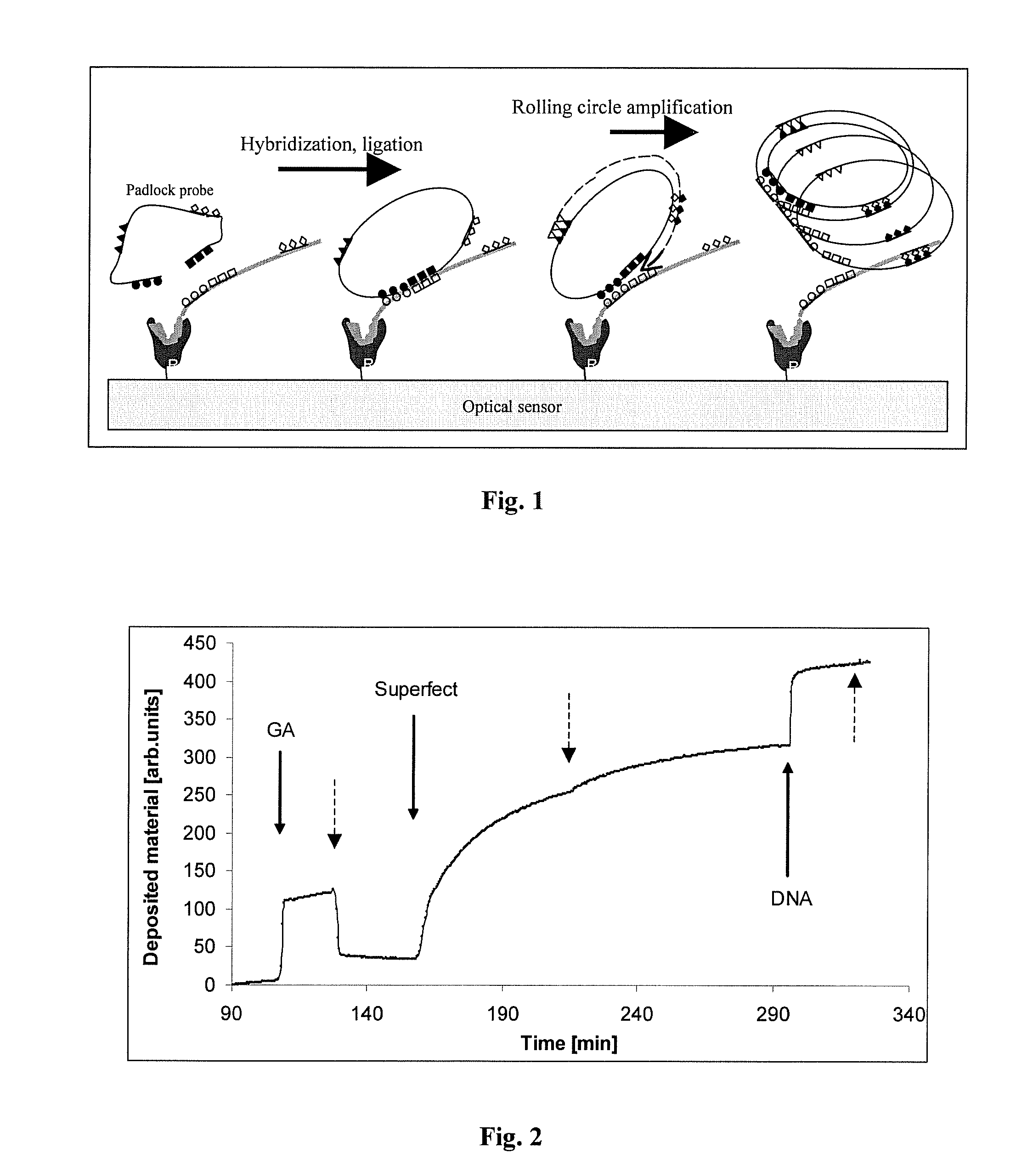
[0026]In accordance with the above, in one embodiment of the present invention, highly selective padlock recognition and rolling circle nucleic acid amplification is adopted to a sensitive optical sensor surface, where the amplified chains are bound and produce real-time optical signals proportional with chain-elongation. The sensitive label-free real-time recording allows for the kinetic analysis of the amplification and the quantification of the initial amount of the captured analyte.
[0034]In order to detect the specific binding of small amount (even 1 molecule) of the ligand, the method of the invention applies an amplifying method, which produces surface-bound material deposition sufficient for detection and being in proportion to the amount of the initially recognized specifically bound analyte and to the advancement of the reaction-time.
[0050]c) contacting said optical sensor surface with a rolling circle amplification (RCA) initiator nucleic acid also comprising a specific binding site for the captured analyte molecules and an anchor sequence ensuring the continuous surface binding of the amplification products in conditions allowing the specific binding of said initiator sequence to said captured analytes;

Problems solved by technology

These methods are time and labor intensive and, therefore, methods were proposed for real time and / or label-free detection of nucleic acids.
However, neither specific teaching nor experimental evidence is put forward to demonstrate the actual elongation of the first polynucleotide, or for the real time detection of the mass change of the first polynucleotide.
Despite the tremendous research and development effort recently put into the development of sensitive, user friendly specific binding assays, and the combination of the existing binding assay arrangements with the convenient label free optical sensor technology, no sensitive optical sensor based binding assay have been developed so far which would be useful for label free real-time detection and quantification of low concentration analytes present in biological specimens.

Method used


Image

  • Label-free optical detection method
  • Label-free optical detection method
  • Label-free optical detection method

Examples


example 1

Functionalization of Optical Sensor Surfaces for DNA Binding

[0071]The surface of a sensor-chip of the optical waveguide light spectroscope (OWLS) had been modified by vacuum sylanization or by traditional APTES-reaction in order to produce abundant —NH2+ reactive groups on the sensor-surface, for further chemical reactions as it is explained in detail below.

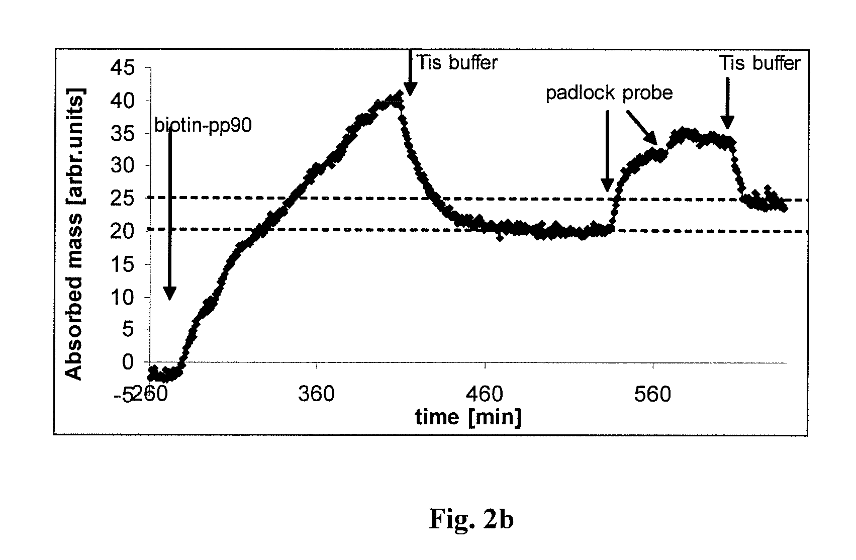
[0072]1.1. The amino-moieties were treated with glutaraldehyde, a bifunctional crosslinking molecule. The resulted glutaraldehyde-coated, reactive surface was reacted with PAMAM dendromeres resulting in a sensor-surface carrying a covalently bound lattice of DNA-binding material. (PAMAM dendromeres are materials used for introducing foreign DNA into cells, because of their strong affinity to DNA and three-dimensional particle nature, which triggers cellular endocytosis ( ).) The building of the “capturing” surface had been followed up by continuous monitoring the material deposition by real-time OWLS measurement (FIG. 2)

[0073]1.2...

example 2

Binding and Elution of DNA on Functionalized Optical Sensor Surfaces

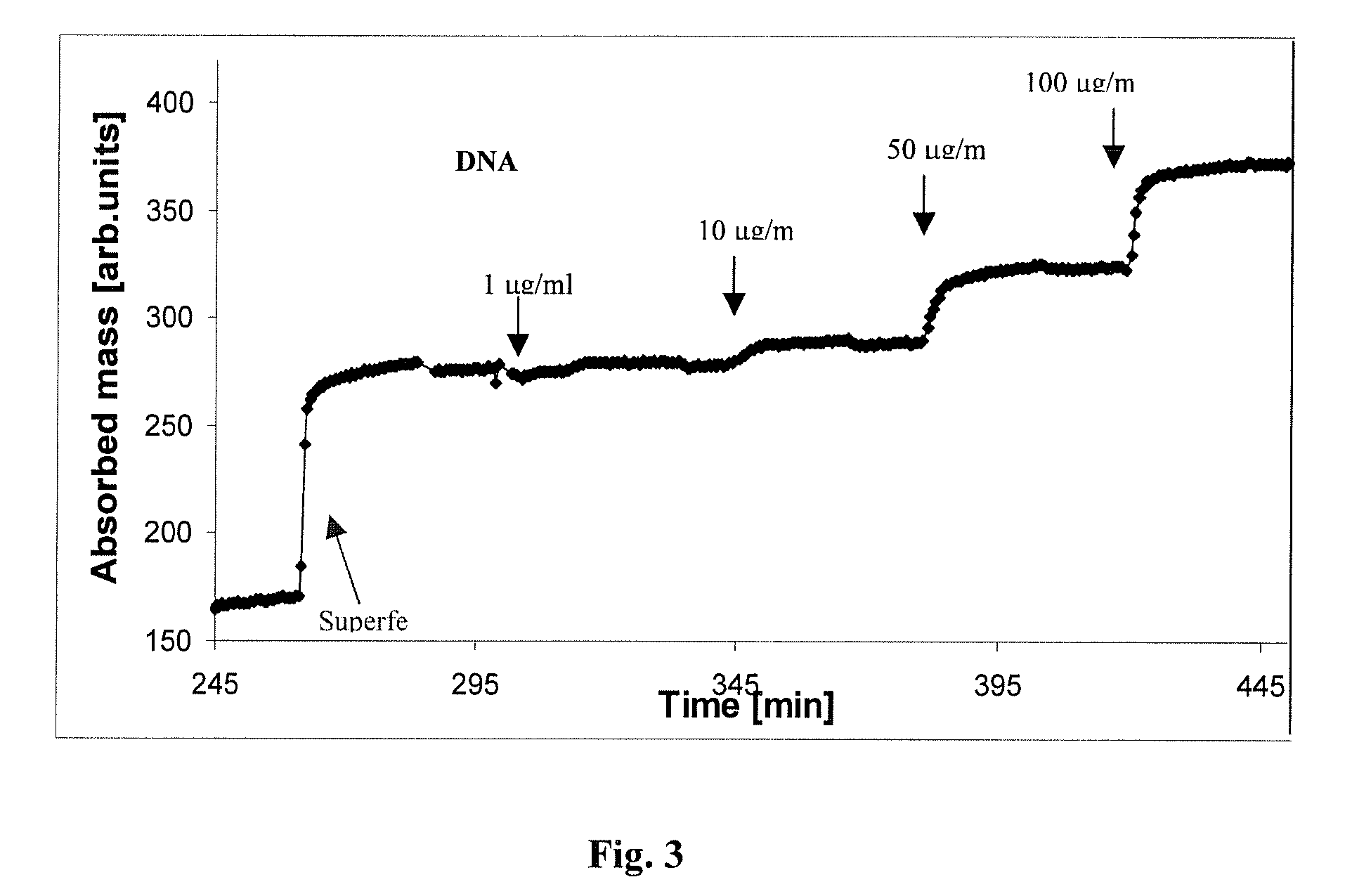
[0078]2.1. The dendromer-functionalized sensor surface captured DNA molecules from a mixture of E.Coli-derived DNA (Sigma) (last step on FIG. 2. and FIG. 3) in a concentration-dependent way (FIG. 3). The stability of DNA-binding was probed by washing with a number of different solutions including Tirs+200 mM Mg2+, SSC, 1.5 M Sodium-acetate, 0.05 M HCl, 2-10 mM EDTA in Tris buffer (TE). None of the eluting solutions could remove the captured DNA.

[0079]The binding could be broken up by washing with low ionic strength solutions at high (>70° C.) temperature (FIG. 3). The conditions for disruption of dendromer-DNA interaction suggest that the binding forces, which stabilize the DNA—dendromer complexes are similar to those coupling DNA double strands together.

[0080]High temperature washing, on the other hand, did not remove the capturing Superfect layer (FIG. 3), thus the functionalized sensors could be used repeatedly.

[...

example 3

Size-Dependent DNA Binding on Sensor Surfaces

[0082]The DNA binding was further analyzed by probing the dendromer-coated sensors with DNA fragments of different size. DNA fractions containing fragments with various lengths were prepared by sonication of the genomic E.Coli DNA (FIG. 4 A).

[0083]50 μg / ml solutions of each fraction were loaded into the OWLS cuvette and the binding was determined under identical conditions. The analyses demonstrated that relatively small DNA fragments used for specific hybridization and molecular recognition can be stably bound to the sensor surface (FIG. 4 B).

[0084]The experimental data demonstrated that relatively small fragments of DNA, those used for specific biomolecular recognition (as in situ hybridization or PCR amplification) can be stably bound to the sensor surface, and the binding can be followed up by real-time optical detection.


PUM

PropertyMeasurementUnit
temperatureaaaaaaaaaa
massaaaaaaaaaa
mass changeaaaaaaaaaa

Abstract

The present provides optical sensor based sensitive, label-free binding assay methods and kits for isothermal real-time detection of the binding of specific analytes (such as nucleic acids, proteins and low molecular weight antigenic or receptor binding ligands) present in low amount in different biological samples. In the binding assays of the invention, the analyte is captured at the specifically pretreated solid surface of optical biosensors in specific recognition reactions (such as hybridization, specific protein-protein interactions and receptor-ligand interactions). The specificity of the methods of the invention is further enhanced by a second specific recognition step using a padlock probe comprising an indicator sequence designed to keep the products of a subsequently performed isothermal nucleic acid amplification method (e.g. rolling circle amplification: RCA) anchored on the sensor surface, enhancing thereby the sensitivity of the detection of the specific binding of the analyte occurred on the sensor surface.

Description

FIELD OF THE INVENTION[0001]The present invention provides optical sensor based sensitive, label-free binding assay methods and kits for isothermal real-time detection of the binding of specific analytes (such as nucleic acids, proteins and low molecular weight antigenic or receptor binding ligands) present in low amount in different biological samples. In the binding assays of the invention, the analyte is captured at the specifically pretreated solid surface of optical biosensors in specific recognition reactions (such as hybridization, specific protein-protein interactions, receptor-ligand interactions, etc). The specificity of the methods of the invention is further enhanced by a second specific recognition step using a padlock probe comprising an indicator sequence designed to keep the products of a subsequently performed isothermal nucleic acid amplification method (e.g. rolling circle amplification: RCA) anchored on the sensor surface, which amplification is used thereby to e...

Claims


Application Information

Patent Timeline
Patent Type & Authority Applications(United States)
IPC IPC(8): C12Q1/68
CPCC12Q1/6825G01N33/54373G01N2458/10C12Q2563/131C12Q2531/125
Inventor SZENDRO, ISTVANMADARASZ, EMILIAERDELYI, KATALINVARGA, BALAZS VIKTOR
Owner SZENDRO ISTVAN
Learn More