
Skin tape stripping: a non-invasive diagnostic strategy for dermal exposure to cytotoxic compounds

a cytotoxic compound and skin tape technology, applied in the direction of immunoglobulins, instruments, peptides, etc., can solve the problems of tissue damage, no single mechanism or clear understanding of the biological damage caused, and the destruction of the us stockpile of mustard chemical warfare agents

Inactive Publication Date: 2009-11-19
US ARMY REPRESENTED BY THE SEC OF THE ARMY
View PDF4 Cites 13 Cited by
  • Summary
  • Abstract
  • Description
  • Claims
  • Application Information

AI Technical Summary

Benefits of technology

[0045]Therefore a need exists for a non-invasive, field expedient str

Problems solved by technology

Mustard agents cause tissue damage within several minutes of contact.
The US stockpile of mustard chemical warfare agents currently is undergoing destruction.
No single mechanism or clear understanding exists for the biological damage caused by mustard agents.
Mustards can destroy a large number of cellular substances by alkylation of DNA, which leads to DNA strand breaks and apoptosis.
Glutathione depletion leads to inactivation of enzymes, loss of calcium homeostasis, lipid peroxidation, cellular membrane breakdown, and cell death.
Unless carried out within 1-2 minutes, decontamination of victims exposed to mustard agents does not prevent subsequent blistering.
No specific treatment or antidote can reverse or prevent the cellular effects of mustard agents.
At this time, there is no definitive diagnostic strategy for determining exposure to HD.
While monoclonal antibodies produced by mammalian hybridoma lines are consistent in their recognition of a single epitope, media and serum costs can make them expensive to produce.
In addition, hybridoma cells only produce murine antibodies and they require specific maintenance procedures for long-term storage.
When performing hybridoma fusions it can be difficult to isolate specific clones that recognize a unique epitope on a bio-threat agent when that marker is rare, sterically shielded, or not immunodominant [2].
Despite its promise, however, phage-antibody technology is not without difficulties.
The last step in particular—expressing the selected antibody genes to make usable quantities of antibody—has proven troublesome, differing idiosyncratically from one antibody to another [2].
This assay format is easily implemented in the laboratory using processed samples, but the challenge is to develop immunoassays that will detect biological agents in the environment.
A drawback of this approach is that quantitative assessments based on color development are subjective unless corrected for time, and analyzed by an automated color densitometer.
Furthermore, these assays are not always as sensitive as other more cumbersome multi-step assays.
However, this kit has some serious drawbacks including a lack of stability of the recombinant Fabs utilized, a lack of sensitivity, a short shelf-live and the need for an automated color densitometer [2].
However, these studies did not allow for investigation of dermal exposure per se since the contaminant was washed from the skin prior to the application of the tape.
Although promising, tape stripping of the stratum corneum for estimation of dermal exposure has been used only with a few compounds of limited interest for occupational health.
Although skin tape peels have been used in dermatological practice and specific investigative study for some time [31], the analysis of samples is typically processed and analyzed in a laboratory using methods such as gas chromatography not readily available in remote locations and requiring heavy use of limited resources and expertise.
Although the practice of skin tape stripping has also been used investigatively to measure the presence of some skin irritants that adhere to the tape and that remain on the skin after tape stripping, such as surface wood coatings, it has yet to be used as a non-invasive diagnostic strategy for skin exposure to chemical and biological agents.
Such a protocol is not only highly time and resource intensive but not conducive to the field expedient requirements that typify the scenario where exposure to sulfur mustard is most common (for example, in an emergency urban setting, or on the battlefield).
However, each of these references suffer from one or more of the following disadvantages: in Benson—the processing of RNA and / or DNA nucleotides in a controlled environment requiring sophisticated laboratory instruments to determine indentify and quantity (RNEASY™ Qiagen, Valencia, Calif. and TRIREAGENT™, Molecular Research Center, Inc., Cincinnati, Ohio); the use of biochips and similar microarrays and conditions necessary to allow for hybridization reactions; and, in Miller—the design is specific to visual inspection in normal light environments and not conducive to immunohistochemical processing and microscopic examination.

Method used


Image

  • Skin tape stripping: a non-invasive diagnostic strategy for dermal exposure to cytotoxic compounds
  • Skin tape stripping: a non-invasive diagnostic strategy for dermal exposure to cytotoxic compounds
  • Skin tape stripping: a non-invasive diagnostic strategy for dermal exposure to cytotoxic compounds

Examples


example 1

Materials and Methods

Human Skin Tape Stripping

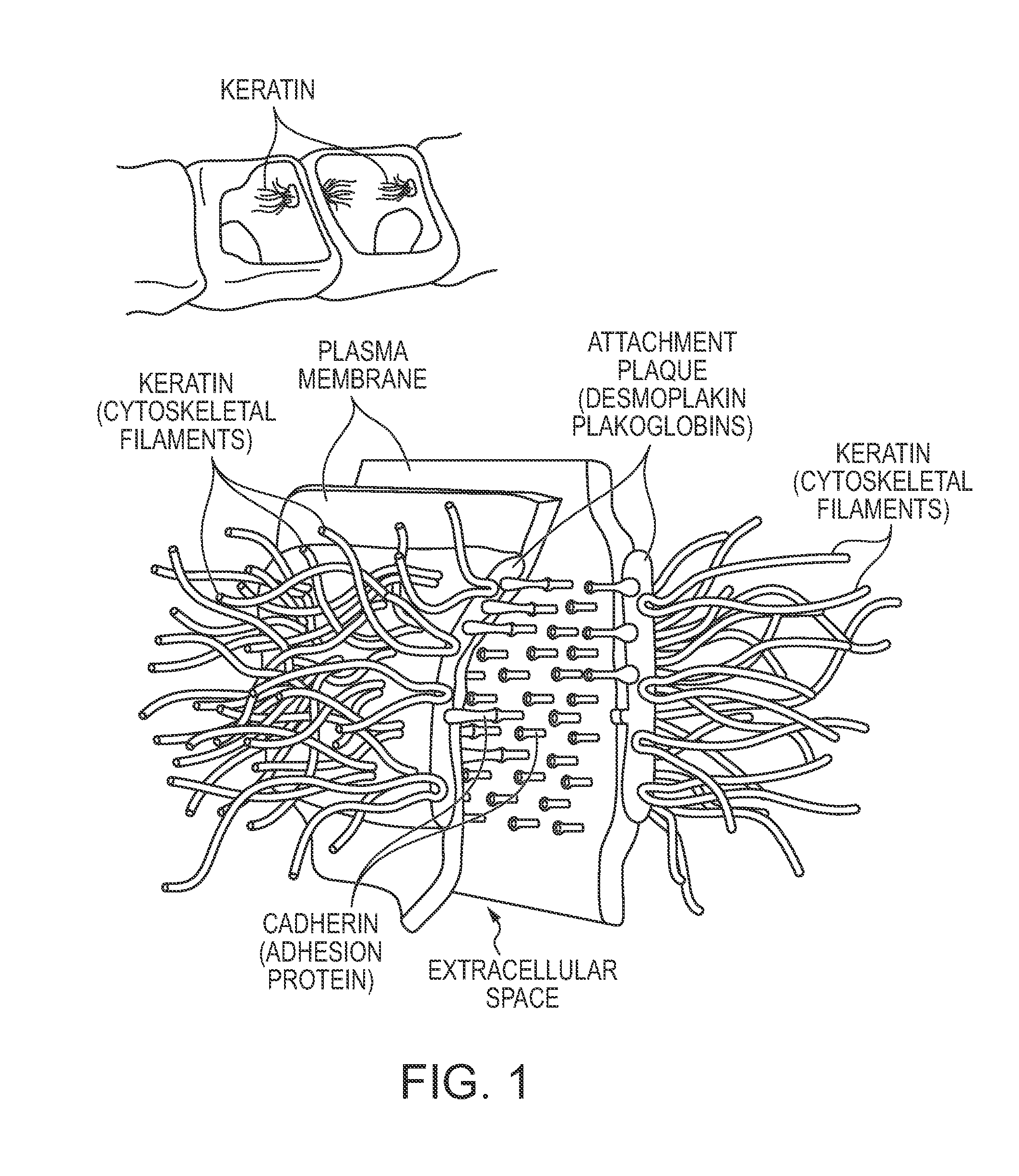
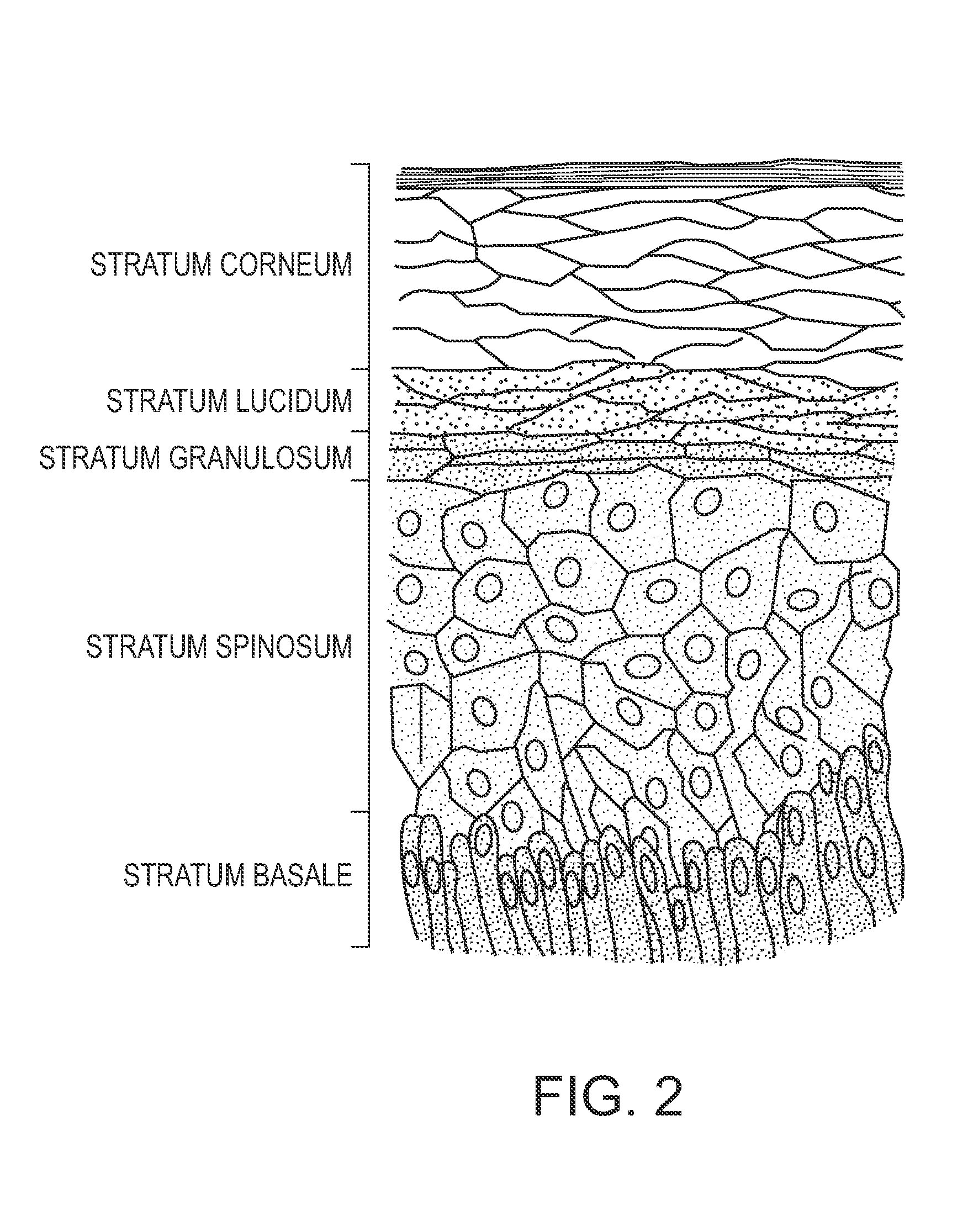
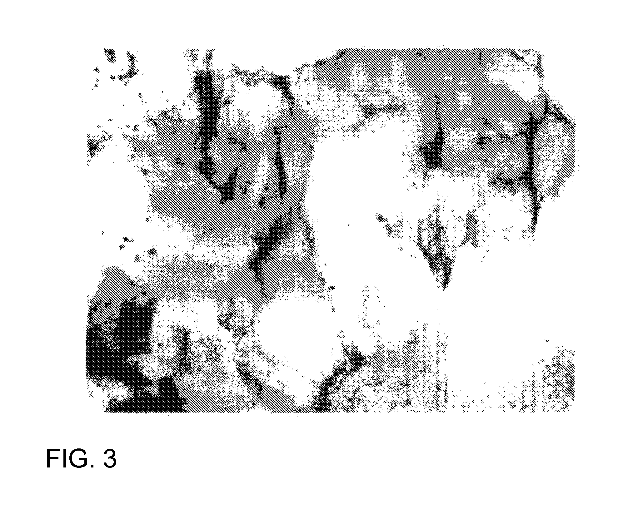
[0090]Double-sided optically clear tapes, Y and Z, and medical grade double-sided tape, A, were purchased from Light Fabrications, Inc. (Rochester, N.Y.). Tapes were cut into one-centimeter squares and adhered to 1′×3′ microscope glass slides; The volar surface of forearms of willing investigators were cleaned with sterile alcohol pads and air dried. Backing from slide tapes was removed and the whole slide pressed. Firmly on the forearm for about 10 seconds. Slides were carefully removed at a 45-degree angle to ensure even cell adherence to the tape's adhesive surface. The slides with attached cells were subjected to different experimental staining and handling paradigms identified below. After each experiment, slides were cover-slipped with permount mounting media and. A 24 mm×30 mm cover glass. Slides were dried overnight and photomicrographed using an OLYMPUS VANOX™ (Olympus Imaging America, Inc., Center Valley, Pa.) light microscope ...

example 2

[0095]Although used for there is not a uniform approach for performing sampling of skin stripping new instrumental procedures are available that provide the means of measuring the physical condition of the skin or for quantifying exogenous or endogenous compounds present within the skin.

[0096]The results of adhesive tape stripping samples were compared using three types of analysis: protein assay of keratin mass and two instrumental approaches using light reflection and pixilation of the digitized image (CUDERM™ Bionet, Inc. Spring, Tex. and VISIOSCAN VC 98™, COURAGE™ and KHAZAKA Koln, Germany).

Methods and Materials

[0097]Adhesive tape strip samples were collected using a standardized technique from paired sites on the palm, wrist and dorsal forearm. One sample was analyzed using the VISIOSCAN VC 98™ for skin flakes that provided total area covered and a calculated desquamation index (D.I.) based on the formula by Schatz et al. [32]. These samples were subsequently analyzed for total...

example 3

Approach 1: Immunization with Keratinaocyte Whole Cell Lysate

[0101]Mustard Gas exposed Keratinocytes Protein Preparation:

[0102]Mustard gas exposed cells—after optimal exposure, wash the cells (three times using sterile PBS) to remove the media components by centrifugation. Finally, suspend the cells in sterile PBS.

[0103]—Adjust cell density to 10 million cells / Ml. Freeze the cells and ship on dry ice. Freeze thaw the cell suspension several times to rupture the cells. Both soluble and insoluble proteins keratinocyte whole cell lysate will be used for the immunization. Measure protein concentration by BCA method.

[0104]Two rabbits will be used for the immunization of keratinocytes whole cell lysate.

Rabbit Protein Protocol, 118 Day:

[0105]

TABLE BDay:Procedure:0NZW or Elite NZW Female Rabbit Pre-bleed(Avg. 5 ml serum)10 ID / SC: 250 μg of protein with FCA21Boost SC: 125 μg with FIA31Test Bleed (Avg. 5 ml serum)32-38ELISA Titer Assay of Bleed*42Boost SC: 125 μg with FIA52Test Bleed (Avg. 5 ...


PUM

PropertyMeasurementUnit
Lengthaaaaaaaaaa
Transparencyaaaaaaaaaa
Cytotoxicityaaaaaaaaaa

Abstract

A non-invasive method for determining a subject's dermal exposure to an agent employing an immunohistochemical procedure on a skin strip applied to an exposed area on the subject's person and detecting agent adduct thereon.

Description

CROSS-REFERENCE TO RELATED APPLICATION[0001]This application claims the benefit of Provisional Application No. 61 / 070,619, filed Mar. 5, 2008; the contents of which are hereby incorporated by reference.RIGHTS[0002]This invention was made with support from the United States Government, specifically, the United States Army Medical Research and Materiel Command, and, accordingly, the United States has certain rights in this invention.FIELD AND BACKGROUND[0003]Mustard agents are vesicants (blister agents) used in warfare to produce casualties, degrade fighting efficiency, and force opposing troops to wear full protective equipment. Mustard agents are cytotoxic alkylating compounds and include nitrogen mustards (HN-1, HN-2, HN-3), sulfur mustards (H, HD, HT), and mustard-lewisite (HL) (abbreviations are NATO designations according to purity grades). Mustard agents are oily liquids ranging from colorless (in pure state) to pale yellow to dark brown, depending on the type and purity. They ...

Claims


Application Information

Patent Timeline
IPC IPC(8): G01N33/567C07K16/00
CPCC07K16/065G01N2800/42G01N2430/00G01N33/6854
Inventor GRAHAM, JOHN S.PETRALI, JOHN P.
Owner US ARMY REPRESENTED BY THE SEC OF THE ARMY
Learn More